 [
[

Scientists study the underlying causes of a disease or test potential new drugs in animal models or cells obtained from human biopsy specimens before clinical trials. However, disease models in animals do not mimic actual human disease, and biopsy specimens are difficult to access for research purposes. Lab-grown tissues engineered from stem cells reprogrammed from human cells, called human induced pluripotent stem cells (hiPSCs), are rapidly emerging as a more reliable and accessible model in medical research.
A study led by Professor Shugo Tohyama and Senior Assistant Professor Hidenori Tani from the Department of Clinical Regenerative Medicine at Fujita Health University in Japan focused on developing robust lab models of cardiomyocytes (CMs), the heart muscle cells. Years of pioneering research have led to the development of high-quality CMs from hiPSCs in a petri dish.
“During efforts to adjust the concentrations of glucose and fatty acids in the culture medium to promote heart tissue maturation, we discovered that excessive fatty acids adversely affected the dilation capacity of the heart tissues,” Tani, the study’s first author, explains regarding this breakthrough.
“Around the same time, a study reported that the administration of high levels of fatty acids and nitric oxide synthase (NOS) inhibitors in mice induced heart failure (HF) with preserved ejection fraction (HFpEF), which led to the conception of this study.”
Building a human HFpEF model
The study, which resulted in the development of a human engineered heart tissue (hEHT) model of HFpEF, was published in Cell Stem Cell.
HFpEF is a defect in the relaxation (diastole) of the heart with normal contraction (systole). It affects more than 30 million people globally but has few effective treatment options and, therefore, poor outcomes.
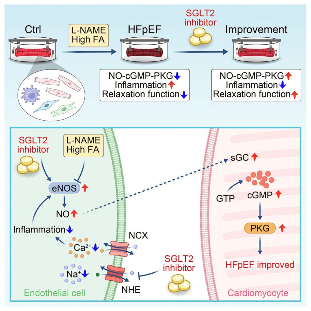
Tohyama and his team induced hiPSC-CMs from readily available hiPSCs, created mature 3D heart tissue using collagen derived from porcine hearts—which the lab had previously developed—and cultured the tissues in vitro using a medium containing high fatty acid levels and L-NG-nitroarginine methyl ester (L-NAME), an NOS inhibitor.
Supporting cells like epicardial cells, endothelial cells (ECs) and macrophages were also added to successfully create an HFpEF-hEHT that structurally and functionally mimicked diastolic HF with normal systole.
Molecular and genetic markers of HFpEF, like increased NT-proBNP and NPPB mRNA levels, reduced expression of key genes related to Ca2+ transport (RYR2, SERCA2A and PLN), increased fibrosis-related genes, and a shift to the stiffer, less pliable type collagen and TITIN, were demonstrated. Increased inflammation was also seen.
Drug testing points to one standout
Using the newly developed HFpEF model, the researchers tested the effectiveness of six drugs currently being used or explored in research for HFpEF treatment. Empagliflozin, a sodium-glucose cotransporter 2 inhibitor (SGLT2i) and antidiabetic medication, was the only drug that could partially prevent the development of diastolic HF.
The combined angiotensin receptor/neprilysin inhibitor, soluble guanylate cyclase activator, phosphodiesterase 5 inhibitor and nicotinamide phosphoribosyltransferase protein activator had no effect on hEHT relaxation time. On the other hand, a hyperpolarization-activated cyclic nucleotide-gated channel inhibitor worsened the disease.
Transcriptomic analysis revealed that SGLT2i had anti-inflammatory effects and partially prevented the increase in inflammatory molecules like IL-1β and IL-6 in HFpEF-hEHT. Also, the presence of ECs was required for SGLT2i to exert its effects. Further studies on the role of supporting cells revealed that the preventive effect of SGLT2i was exerted through ECs.
Inhibition of the eNOS-NO-cGMP-PKG signaling pathway in ECs was found to be the main pathological mechanism of HFpEF. SGLT2i was found to exert its anti-inflammatory effects by partially removing this inhibition and by improving the removal of Na+ and Ca2+ ions from the cells through their exchanger channels, thereby preventing their accumulation in the tissues.
A platform for tailored therapies
“The human HFpEF model established in this study can be applied to uncovering the pathophysiology of HFpEF and to research on aging,” Tohyama says.
“Furthermore, it is expected to serve as a research foundation that will contribute to the development of new therapeutic agents, the evaluation of drug efficacy, and personalized medicine for individual patients, paving the way for further research in the future,” Tohyama adds.
Publication details
Hidenori Tani et al, Human iPSC-derived engineered heart tissue model of diastolic dysfunction in heart failure with preserved ejection fraction, Cell Stem Cell (2026). DOI: 10.1016/j.stem.2026.06.007
Journal information:
Cell Stem Cell
Key medical concepts
Clinical categories


Citation:
Lab-grown tissue reveals how inflammation may drive heart relaxation failure (2026, September 2)
retrieved 2 September 2026
from https://medicalxpress.com/news/2026-09-lab-grown-tissue-reveals-inflammation.html
This document is subject to copyright. Apart from any fair dealing for the purpose of private study or research, no
part may be reproduced without the written permission. The content is provided for information purposes only.

Khamrah by Lattafa for Men - 3.4 oz EDP Spray
4% Off
Ghost Sweetheart Eau de Toilette | Pineapple, Jasmine and Sandalwood | Perfume for Women 50 ml
50% Off
Marc Jacobs Dot Eau De Parfum for Women, 100 ml
42% Off
Ted Baker W Eau de Toilette for Her, Fig Leaf, White Peony and Violet Top Notes, Pink Orchid and Raspberry Middle Notes, 75ml
£11.77 (£15.69 / 100 ml) (as of 04/09/2026 04:57 GMT +01:00 - More infoProduct prices and availability are accurate as of the date/time indicated and are subject to change. Any price and availability information displayed on [relevant Amazon Site(s), as applicable] at the time of purchase will apply to the purchase of this product.)
Ted Baker Woman Pink Eau de Toilette Spray Floral Green Feminine Fragrance, Opening Notes are Fresh Peach, Bergamot and Tangerine with Warm Musk, Vanilla and Vetiver Base, 100ml
11% Off
Vera Wang Princess Eau de Toilette - 30 ml

Choco Musk 50ml Eau De Parfum for men and women | Chocolate Musk by Jannat Aromas
17% Off
Christina Aguilera Signature Eau de Parfum (50ml) Floral, Fruity & Exotic Scent, Luxury Fragrance for Women
9% Off
Calvin Klein - Eau De Toilette CKIN2U - Calvin Klein Women, Ladies Perfume, Women's Perfume, Calvin Klein Perfume, Calvin Klein One - 150 ml
5% Off
Jimmy Choo Flash Eau de Parfum, 60 ml (Pack of 1)
3% Off
Fruit of the Loom Men's Heavy T Shirt, White, XL UK
28% Off
ATNKE LED Lighted Beanie Cap,USB Rechargeable Running Hat Ultra Bright 4 LED Waterproof Light Winter Warm Gifts for Men and Women/Pink
17% Off
Men's 1/4 Zip Pullover UK Sale Clearance, Fleece Sweatshirt Casual Jumper Long Sleeve T-shirt Top Stand Collar Sweater Plain Pullover Sports Leisure Workwear Quarter Zip Sweater Lightweight Jumpers
£5.88 (as of 12/11/2025 00:52 GMT +01:00 - More infoProduct prices and availability are accurate as of the date/time indicated and are subject to change. Any price and availability information displayed on [relevant Amazon Site(s), as applicable] at the time of purchase will apply to the purchase of this product.)
Crevice Cleaning Brush, Bathroom Tile Groove Gap Cleaning Brush,Premium Crevice Cleaning Tool Aluminum Support with 15° Angle Magic Brush, Thin Brush for Home Kitchen
19% Off
Wireless Earbuds, Bluetooth 5.3 Headphones in Ear with HiFi Stereo Deep Bass, 4 ENC Noise Cancelling Mic Wireless Earphones 40H Playtime, Bluetooth Earbuds Dual LED Display, IP7 Waterproof, USB-C
42% Off