[
[
Soft electrodes designed to perfectly match a person’s brain surface may help advance neural interfaces for neurodegenerative disease monitoring and treatment, according to a new study led by Penn State researchers. Neural interfaces are powered by tiny sensors capable of tracking biophysical signals, known as bioelectrodes. These sensors are usually made from stiff materials in a one-size-fits-all design that struggles to match the brain’s complex structure. The researchers have created a novel approach to 3D printing bioelectrodes that can stretch and morph to fit the minor differences that make every brain unique.
Simulating unique brain structures
The team used software to simulate detailed brains based on MRI scans taken from 21 human patients, shaping a set of electrodes tailored for brains’ specific structures before 3D printing the electrodes and models of the brains. In a paper published in Advanced Materials, they reported that their electrodes better fit the structure of the brain than traditional designs, while remaining effective and biologically compatible, even in tests done in rats.
The folds in the human brain are created through a process known as gyrification, where the cortical sheet on the outer wall of the brain bunches up into ridges, known as gyri, and grooves, known as sulci. This helps cells across the brain communicate at high speeds, and allows for a relatively large organ to fit compactly in the skull—a spread-out adult brain would be around 2,000 square centimeters, or about the size of two large pizzas.
Why one-size-fits-all falls short
Although the major cortical folds are consistent across individuals, the precise layout of the brain’s gyri and sulci changes substantially from person to person, according to Tao Zhou, Wormley Family Early Career Professor, assistant professor of engineering science and mechanics and corresponding author on the paper. However, traditional bioelectrode designs don’t take this into account.
“Each person has a different brain structure, depending on their height, weight, age, sex and more,” said Zhou, who also holds an affiliation in biomedical engineering and the center for neural engineering at Penn State. “Despite this, we try to fit neural interfaces onto brains like they have identical structures. This motivated us to create electrodes that are tailored for each individual, based on the structure of their brain.”
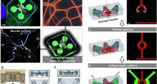
Hydrogel and honeycomb design
The electrodes are built mainly from a water-rich material known as hydrogel to better match the soft tissues and patient-specific geometry of a brain. Furthermore, the team used a novel honeycomb-inspired structure that offers flexibility and strength, while remaining cost-effective and quick to print, according to Zhou.
“The honeycomb structure helps us significantly reduce the stiffness of the electrodes, without sacrificing their mechanical strength,” Zhou said. “What’s more, the structure helps us reduce the overall material used during fabrication, reducing production time, cost and environmental impact.”
From MRI scan to 3D-printed match
Production starts by taking an MRI scan of a patient’s brain, which is used to conduct finite element analysis—a process that creates a detailed simulation of a person’s neural structure. This analysis is then rendered as a 3D model of the patient’s brain, where the team uses computer software to tailor a bioelectrode specifically morphed to fit the ridges and grooves of the cerebral cortex.
After shaping, the team 3D prints the hydrogel electrode using direct ink printing, a technique that can create electrodes capable of monitoring and transmitting brain signals over a relatively small surface. For this study, the team 3D printed models of 21 different participant brains, applying their electrodes and physically measuring how accurately the electrodes could fit the brain surface. Zhou explained how traditional fabrication approaches require specialized facilities like clean rooms, making them incredibly expensive to customize—3D printing allows the team to personalize and manufacture electrodes much faster, for a fraction of the price.
Softer contact, stronger brain signals
Compared to traditional approaches, the hydrogel-based electrodes follow the structure of the brain more precisely. Zhou said their approach produces electrodes that exhibit nearly perfect connectivity to electrical signals present in the brain. Additionally, because the stretchy gel is so malleable, it can be applied to the soft brain tissue without causing damage, compared to the stiff materials comprising other designs that could damage tissue.
According to Zhou, the softness of their electrodes enables closer and more stable contact with the brain, in turn facilitating higher-quality, more reliable monitoring. Moreover, bioelectrodes made with this approach don’t impact fluid transport around the brain, a critical aspect of brain function that many traditional electrodes disrupt.
“Personalizing the electrodes to the brain’s specific structure substantially improves their reliability,” Zhou said. “Because they conform to the brain better, the signal quality itself is significantly improved.”
Testing in rats and future use
To further study their electrodes, the team placed them onto the brains of rat models over a period of 28 days. The rats did not exhibit any immune response to the printed electrodes, a key consideration in biodevice development, Zhou said. Additionally, the electrodes did not exhibit performance degradation, while offering sensitive and accurate readings of the electric and physiological signals in the brain.
Zhou said he believes that this printing method could serve as a framework for the commercial-scale printing of bioelectrodes customized for specific patients. Although these systems are traditionally used for monitoring neural activity, the team plans to explore how personalized electrodes may contribute to neurological treatments.
“We are looking to further improve this technology to optimize the electrodes to monitor for specific diseases,” Zhou said. “In the future, we would really like to work with patients to see how this approach could support brain monitoring and disease treatment in clinical settings.”
Publication details
Marzia Momin et al, 3D‐Printable, Honeycomb‐Inspired Tissue‐Like Bioelectrodes for Patient‐Specific Neural Interface, Advanced Materials (2026). DOI: 10.1002/adma.202516291
Journal information:
Advanced Materials
Key medical concepts
Hydrogel, Polyethylene Glycol DimethacrylateFinite Element Analysis
Clinical categories
Citation:
3D-printed brain sensors may unlock personalized neural monitoring (2026, April 18)
retrieved 18 April 2026
from https://medicalxpress.com/news/2026-04-3d-brain-sensors-personalized-neural.html
This document is subject to copyright. Apart from any fair dealing for the purpose of private study or research, no
part may be reproduced without the written permission. The content is provided for information purposes only.

Khamrah by Lattafa for Men - 3.4 oz EDP Spray
4% Off
Ghost Sweetheart Eau de Toilette | Pineapple, Jasmine and Sandalwood | Perfume for Women 50 ml
50% Off
Marc Jacobs Dot Eau De Parfum for Women, 100 ml
42% Off
Ted Baker W Eau de Toilette for Her, Fig Leaf, White Peony and Violet Top Notes, Pink Orchid and Raspberry Middle Notes, 75ml
£11.77 (£15.69 / 100 ml) (as of 18/04/2026 02:58 GMT +01:00 - More infoProduct prices and availability are accurate as of the date/time indicated and are subject to change. Any price and availability information displayed on [relevant Amazon Site(s), as applicable] at the time of purchase will apply to the purchase of this product.)
Ted Baker Woman Pink Eau de Toilette Spray Floral Green Feminine Fragrance, Opening Notes are Fresh Peach, Bergamot and Tangerine with Warm Musk, Vanilla and Vetiver Base, 100ml
11% Off
Vera Wang Princess Eau de Toilette - 30 ml

Choco Musk 50ml Eau De Parfum for men and women | Chocolate Musk by Jannat Aromas
17% Off
Christina Aguilera Signature Eau de Parfum (50ml) Floral, Fruity & Exotic Scent, Luxury Fragrance for Women
9% Off
Calvin Klein - Eau De Toilette CKIN2U - Calvin Klein Women, Ladies Perfume, Women's Perfume, Calvin Klein Perfume, Calvin Klein One - 150 ml
5% Off
Jimmy Choo Flash Eau de Parfum, 60 ml (Pack of 1)
3% Off
Fruit of the Loom Men's Heavy T Shirt, White, XL UK
28% Off
ATNKE LED Lighted Beanie Cap,USB Rechargeable Running Hat Ultra Bright 4 LED Waterproof Light Winter Warm Gifts for Men and Women/Pink
17% Off
Men's 1/4 Zip Pullover UK Sale Clearance, Fleece Sweatshirt Casual Jumper Long Sleeve T-shirt Top Stand Collar Sweater Plain Pullover Sports Leisure Workwear Quarter Zip Sweater Lightweight Jumpers
£5.88 (as of 12/11/2025 00:52 GMT +01:00 - More infoProduct prices and availability are accurate as of the date/time indicated and are subject to change. Any price and availability information displayed on [relevant Amazon Site(s), as applicable] at the time of purchase will apply to the purchase of this product.)
Crevice Cleaning Brush, Bathroom Tile Groove Gap Cleaning Brush,Premium Crevice Cleaning Tool Aluminum Support with 15° Angle Magic Brush, Thin Brush for Home Kitchen
19% Off
Wireless Earbuds, Bluetooth 5.3 Headphones in Ear with HiFi Stereo Deep Bass, 4 ENC Noise Cancelling Mic Wireless Earphones 40H Playtime, Bluetooth Earbuds Dual LED Display, IP7 Waterproof, USB-C
42% Off