 [
[

Early diagnosis and noninvasive monitoring of neurological disorders require sensitivity to elusive cellular-level alterations that emerge much earlier than volumetric changes observable with millimeter-resolution medical imaging.
Morphological changes in axons—the tube-like projections of neurons that transmit electrical signals and constitute the bulk of the brain’s white matter—are a common hallmark of a wide range of neurological disorders, as well as normal development and aging.
A study from the University of Eastern Finland (UEF) and the New York University (NYU) Grossman School of Medicine establishes a direct analytical link between the axonal microgeometry and noninvasive, millimeter-scale diffusion MRI (dMRI) signals—diffusion MRI measures the diffusion of water molecules within biological tissues and is sensitive to tissue microstructure.
The study is published in the journal Nature Communications.
Advances in axonal imaging and analysis
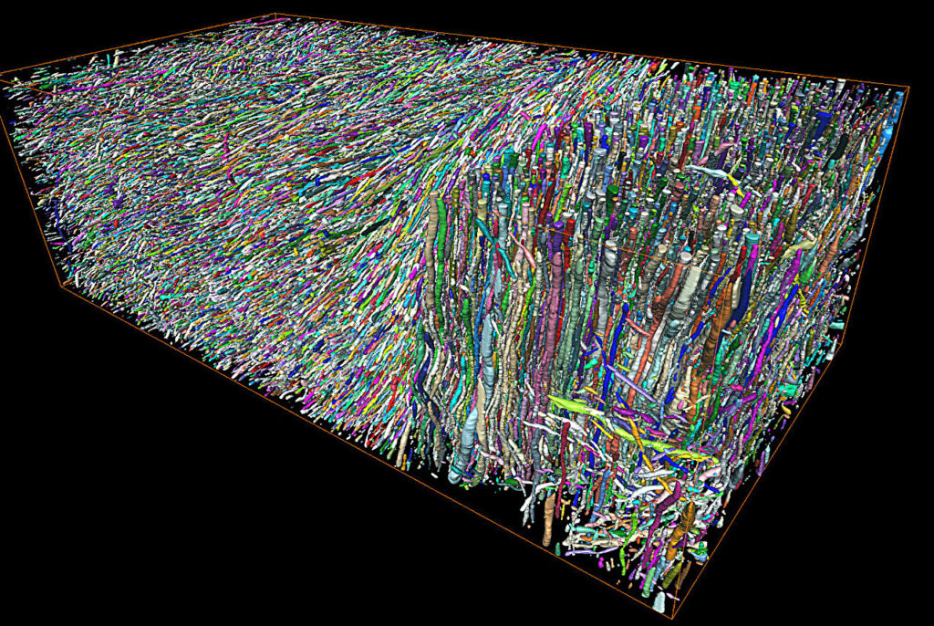
The discovery by Academy Research Fellow Dr. Ali Abdollahzadeh at UEF and Professor Dmitry S. Novikov at NYU emerged from the convergence of two advances: unprecedented three-dimensional reconstruction of white matter microstructure using large-scale volume electron microscopy (vEM) and modern theoretical developments in diffusion physics.
Advanced computational methods developed by Abdollahzadeh made it possible to reconstruct hundreds of thousands of axons with nanometer precision from vEM images acquired in Research Director Alejandra Sierra’s lab at UEF, establishing what is now the world’s largest quantitative three-dimensional reference of white matter microgeometry.
“These reconstructions revealed how axons deviate from simple straight tubes. We directly measured fluctuations in axonal cross-section and undulations along their length, among other morphological parameters,” says Abdollahzadeh.
New framework for diffusion MRI
During Abdollahzadeh’s postdoctoral research at NYU, led by Professors Dmitry S. Novikov and Els Fieremans, and building on Novikov’s pioneering advances in diffusion physics, Abdollahzadeh and Novikov developed a scattering framework for diffusion in cross-sectionally varying axons, yielding an exact solution to the Fick–Jacobs equation governing axial diffusion along axons.
“To solve a complex physical problem means identifying which parameters truly matter. Despite the infinite geometric complexity of axons, we found that only two structural parameters govern axial diffusion at experimentally accessible diffusion times: the average reciprocal cross-section and the variance of long-range cross-sectional fluctuations,” says Novikov.
Experimental validation and future directions
Validation in an experimental rat model of traumatic brain injury at UEF’s Kuopio Biomedical Imaging Unit showed that ex vivo dMRI is sensitive to these microstructural parameters, even months after injury, transforming the interpretation of dMRI into a quantitative probe of axonal geometry.
These findings open new avenues for noninvasive biomarkers of white matter injury. Axonal shape changes are a hallmark of many neurological disorders, and this advance now enables their detection, monitoring and assessment of treatment response using dMRI.
Looking ahead, the team is extending this framework to animal models of diverse neurological pathologies at the Kuopio Biomedical Imaging Unit, headed by Professor Olli Gröhn, using the newly installed state-of-the-art 9.4-T MRI system.
In parallel, translation to the human brain is advancing on human MRI scanners at Kuopio University Hospital and at NYU with the newly installed next-generation Connectom.X scanner with ultra-strong gradients.
“The combination of MRI in animal models and clinical MRI creates a translational path from nanometer-scale tissue microstructure to human neuroimaging, allowing us to test such microstructure-specific biomarkers in patients for the first time,” says Fieremans.
Publication details
Ali Abdollahzadeh et al, Scattering approach to diffusion quantifies axonal damage in brain injury, Nature Communications (2025). DOI: 10.1038/s41467-025-64793-1
Journal information:
Nature Communications
Clinical categories
Citation:
Turning MRI into a quantitative microscope to detect white matter injury (2026, January 19)
retrieved 19 January 2026
from https://medicalxpress.com/news/2026-01-mri-quantitative-microscope-white-injury.html
This document is subject to copyright. Apart from any fair dealing for the purpose of private study or research, no
part may be reproduced without the written permission. The content is provided for information purposes only.

Khamrah by Lattafa for Men - 3.4 oz EDP Spray
4% Off
Ghost Sweetheart Eau de Toilette | Pineapple, Jasmine and Sandalwood | Perfume for Women 50 ml
50% Off
Marc Jacobs Dot Eau De Parfum for Women, 100 ml
42% Off
Ted Baker W Eau de Toilette for Her, Fig Leaf, White Peony and Violet Top Notes, Pink Orchid and Raspberry Middle Notes, 75ml
£11.77 (£15.69 / 100 ml) (as of 20/06/2026 03:33 GMT +01:00 - More infoProduct prices and availability are accurate as of the date/time indicated and are subject to change. Any price and availability information displayed on [relevant Amazon Site(s), as applicable] at the time of purchase will apply to the purchase of this product.)
Ted Baker Woman Pink Eau de Toilette Spray Floral Green Feminine Fragrance, Opening Notes are Fresh Peach, Bergamot and Tangerine with Warm Musk, Vanilla and Vetiver Base, 100ml
11% Off
Vera Wang Princess Eau de Toilette - 30 ml

Choco Musk 50ml Eau De Parfum for men and women | Chocolate Musk by Jannat Aromas
17% Off
Christina Aguilera Signature Eau de Parfum (50ml) Floral, Fruity & Exotic Scent, Luxury Fragrance for Women
9% Off
Calvin Klein - Eau De Toilette CKIN2U - Calvin Klein Women, Ladies Perfume, Women's Perfume, Calvin Klein Perfume, Calvin Klein One - 150 ml
5% Off
Jimmy Choo Flash Eau de Parfum, 60 ml (Pack of 1)
3% Off
Fruit of the Loom Men's Heavy T Shirt, White, XL UK
28% Off
ATNKE LED Lighted Beanie Cap,USB Rechargeable Running Hat Ultra Bright 4 LED Waterproof Light Winter Warm Gifts for Men and Women/Pink
17% Off
Men's 1/4 Zip Pullover UK Sale Clearance, Fleece Sweatshirt Casual Jumper Long Sleeve T-shirt Top Stand Collar Sweater Plain Pullover Sports Leisure Workwear Quarter Zip Sweater Lightweight Jumpers
£5.88 (as of 12/11/2025 00:52 GMT +01:00 - More infoProduct prices and availability are accurate as of the date/time indicated and are subject to change. Any price and availability information displayed on [relevant Amazon Site(s), as applicable] at the time of purchase will apply to the purchase of this product.)
Crevice Cleaning Brush, Bathroom Tile Groove Gap Cleaning Brush,Premium Crevice Cleaning Tool Aluminum Support with 15° Angle Magic Brush, Thin Brush for Home Kitchen
19% Off
Wireless Earbuds, Bluetooth 5.3 Headphones in Ear with HiFi Stereo Deep Bass, 4 ENC Noise Cancelling Mic Wireless Earphones 40H Playtime, Bluetooth Earbuds Dual LED Display, IP7 Waterproof, USB-C
42% Off