[
[
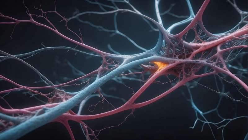
A new study published in Nature Communications shows that human glial progenitor cells are a promising and safe cell product for transplantation. The research also defines the transcriptional and epigenetic signatures of these cells as they mature into astrocytes and oligodendrocytes, two essential support cell types in the brain.
“We believe that glia are particularly divergent compared to other species,” said lead study author John Mariani, Ph.D., a neuroscientist with University of Rochester Medicine and the first author of the new paper.
“For this study, we grow these cells in vitro, and then we transplant them. We wanted to know what they look like before we transplant them and what they look like after. This sets the stage for long-term manipulation of these cells to engraft better, to respond better to the cues, and understand this process better, since it is an approach we are pursuing for cell therapies.”
This transplantation model may have implications for disorders such as multiple sclerosis, leukodystrophies, and Huntington’s disease, in which myelination, the insulating process that helps nerve cells communicate efficiently, is disrupted.
Glial progenitor cells as a cell therapy
The loss of functional astrocytes and oligodendrocytes can lead to a variety of neurological disorders. Glial progenitor cells (GPCs) are precursor cells that can differentiate into mature astrocytes and oligodendrocytes. Because of this potential, GPCs have been studied as a potential cell therapy for glial diseases, and Steve Goldman, MD, Ph.D., and his lab have long been leaders in this area.
Transplanting pluripotent stem cells directly into mice or humans can be dangerous, causing a teratoma to form, a benign tumor that can cause severe neurological complications by occupying critical space in the brain.
In the new study, the GPCs showed no pluripotent signatures in culture, and mature identity persisted after transplantation into mice, indicating that the cells were highly specified, functional, and safe.
A transcriptional and epigenetic map
Researchers began by growing GPCs in vitro and used single-cell RNA and ATAC sequencing to create a comprehensive, genome-wide molecular blueprint of these cells. They found that the GPCs in culture were mature, as evidenced by their lack of pluripotent markers and by remodeling of gene expression as the cells transitioned into specific cell types, thereby activating the cells’ proper function.
The researchers then transplanted the cells into a chimeric, immunodeficient, myelin-deficient mouse model developed in Goldman’s lab, placing the cells in the corpus callosum, the brain’s largest white matter structure. The human glial cells eventually outcompeted and replaced the dysfunctional mouse glial cells and began remyelinating the brain.
Remarkably, the human GPCs extracted back from the mice after engraftment showed a pronounced shift in identity. The GPCs showed strong differentiation and mature astrocyte and oligodendrocyte cell profiles, suggesting that “the cues in the mouse are very good at tightening these cells up to behave,” explained Mariani.
Within the tissue microenvironment, the researchers also identified genes, pathways, and networks that may be responsible for the maturation of the cells to astrocytes and oligodendrocytes.
These networks have strong implications for future studies aimed at understanding how cells respond post-transplantation and how to modulate these cells for the treatment of glial diseases.
Publication details
John N. Mariani et al, Charting the transition from in vitro gliogenesis to the in vivo maturation of human glial progenitor cells transplanted into the hypomyelinated mouse brain, Nature Communications (2026). DOI: 10.1038/s41467-026-71803-3
Journal information:
Nature Communications
Clinical categories
Who’s behind this story?

Sadie Harley

Citation:
From dish to brain: Researchers chart human glial cell maturation (2026, May 29)
retrieved 29 May 2026
from https://medicalxpress.com/news/2026-05-dish-brain-human-glial-cell.html
This document is subject to copyright. Apart from any fair dealing for the purpose of private study or research, no
part may be reproduced without the written permission. The content is provided for information purposes only.

Khamrah by Lattafa for Men - 3.4 oz EDP Spray
4% Off
Ghost Sweetheart Eau de Toilette | Pineapple, Jasmine and Sandalwood | Perfume for Women 50 ml
50% Off
Marc Jacobs Dot Eau De Parfum for Women, 100 ml
42% Off
Ted Baker W Eau de Toilette for Her, Fig Leaf, White Peony and Violet Top Notes, Pink Orchid and Raspberry Middle Notes, 75ml
£11.77 (£15.69 / 100 ml) (as of 06/09/2026 04:59 GMT +01:00 - More infoProduct prices and availability are accurate as of the date/time indicated and are subject to change. Any price and availability information displayed on [relevant Amazon Site(s), as applicable] at the time of purchase will apply to the purchase of this product.)
Ted Baker Woman Pink Eau de Toilette Spray Floral Green Feminine Fragrance, Opening Notes are Fresh Peach, Bergamot and Tangerine with Warm Musk, Vanilla and Vetiver Base, 100ml
11% Off
Vera Wang Princess Eau de Toilette - 30 ml

Choco Musk 50ml Eau De Parfum for men and women | Chocolate Musk by Jannat Aromas
17% Off
Christina Aguilera Signature Eau de Parfum (50ml) Floral, Fruity & Exotic Scent, Luxury Fragrance for Women
9% Off
Calvin Klein - Eau De Toilette CKIN2U - Calvin Klein Women, Ladies Perfume, Women's Perfume, Calvin Klein Perfume, Calvin Klein One - 150 ml
5% Off
Jimmy Choo Flash Eau de Parfum, 60 ml (Pack of 1)
3% Off
Fruit of the Loom Men's Heavy T Shirt, White, XL UK
28% Off
ATNKE LED Lighted Beanie Cap,USB Rechargeable Running Hat Ultra Bright 4 LED Waterproof Light Winter Warm Gifts for Men and Women/Pink
17% Off
Men's 1/4 Zip Pullover UK Sale Clearance, Fleece Sweatshirt Casual Jumper Long Sleeve T-shirt Top Stand Collar Sweater Plain Pullover Sports Leisure Workwear Quarter Zip Sweater Lightweight Jumpers
£5.88 (as of 12/11/2025 00:52 GMT +01:00 - More infoProduct prices and availability are accurate as of the date/time indicated and are subject to change. Any price and availability information displayed on [relevant Amazon Site(s), as applicable] at the time of purchase will apply to the purchase of this product.)
Crevice Cleaning Brush, Bathroom Tile Groove Gap Cleaning Brush,Premium Crevice Cleaning Tool Aluminum Support with 15° Angle Magic Brush, Thin Brush for Home Kitchen
19% Off
Wireless Earbuds, Bluetooth 5.3 Headphones in Ear with HiFi Stereo Deep Bass, 4 ENC Noise Cancelling Mic Wireless Earphones 40H Playtime, Bluetooth Earbuds Dual LED Display, IP7 Waterproof, USB-C
42% Off