 [
[


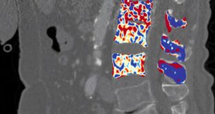
A research team led by Lawrence Berkeley National Laboratory (Berkeley Lab) has developed a new imaging technique that could allow modern medical scanners to image targeted alpha therapy (TAT), an emerging cancer therapy for the diagnosis and treatment of cancer. The advance could enable the widespread adoption of this new cancer therapy.
Targeted cancer therapies use specially engineered radioisotopes that target and kill cancer cells. One of the most promising of these new approaches uses alpha-emitting radioisotopes such as actinium-225. Clinicians still lack a reliable way to image actinium-225 because the medical scanners available today lack the resolution and detection efficiency needed to detect the activity of alpha-emitting radioisotopes in the body. But now a new technique called TOF-CGI (time-of-flight cascade gamma-ray imaging) could close that gap. The work was recently published in the Journal of Nuclear Medicine.
“TOF-CGI brings us a step closer to helping clinicians diagnose and treat cancer in a way that wasn’t possible before,” said Javier Caravaca, a staff scientist in Berkeley Lab’s Nuclear Science Division and principal investigator of the new study.

Advancing TAT imaging with TOF-CGI
Targeted radioisotopes are engineered with targeting molecules to form a radioactive compound. The targeting molecule guides this compound to a cancer cell, where it binds. Actinium-225 is a leading candidate for targeted alpha therapy because of its exceptional potency, and its half-life of around 10 days is ideal for localized treatment with fewer side effects. Actinium-225 emits four alpha particles as it decays, splintering a cancer cell’s DNA into fragments without harming nearby healthy cells. This prevents the cancer cell from regenerating and slows the progression of disease.
Actinium-225 could also be a powerful imaging agent. As the radioisotope decays, its daughter products release gamma photon pairs; the idea is to use these signals to locate tumors while tracking how they respond to treatment over time. Advanced algorithms allow medical scanners to reassemble such data points into 3D images of a tumor.
“Traditionally, there have been only two ways to image cancer: SPECT (single-photon emission computed tomography) or PET (positron emission tomography). Although these techniques have led to big advances in cancer imaging, neither has so far met the demanding requirements to image targeted alpha therapy accurately. Our new study is the first to show that there could be a third way to image cancer with the alpha therapy radioisotope actinium-225,” Caravaca said.

SPECT scanners are commonly used in hospitals, but they lack the required sensitivity to accurately generate images from the detected gamma rays in targeted alpha therapy. SPECT uses thick, heavy metal shields (collimators) to detect single gamma rays—but this only works well when the flux of gamma rays is large. And because targeted alpha therapy is so powerful, only small doses of the radioisotope—about 100 times less than a conventional radiotracer—are administered to patients. Altogether, these constraints add up to low gamma-ray sensitivity and noisy images.
On the other hand, PET promises higher gamma-ray sensitivity and image quality because it doesn’t use collimators. PET works by picking up “positron annihilation” events—when a positron collides with an electron, two gamma-ray photons are emitted back-to-back. The PET algorithm quickly combines tens of thousands of these data points to generate an image of the radioisotope accumulation in the body, such as in a tumor.
Researchers have long assumed that PET scanners wouldn’t work with actinium-225 because the radioisotope emits alpha particles, not positrons. “But one day we wondered, what if we could exploit actinium-225’s gamma-ray pairs to demonstrate targeted alpha therapy in a PET scanner?”
So Caravaca wrote a new algorithm from scratch. TOF-CGI is an image reconstruction algorithm that uses the detected gamma-ray pairs to pinpoint where actinium-225 decayed and reconstruct that data into a 3D image of actinium-225 in the body. TAT emits thousands of signals as the radionuclide decays, so leveraging the GPU computation capabilities at the National Energy Research Scientific Computing Center (NERSC) was essential to significantly accelerate the image-generation process during the development and testing of this technique.
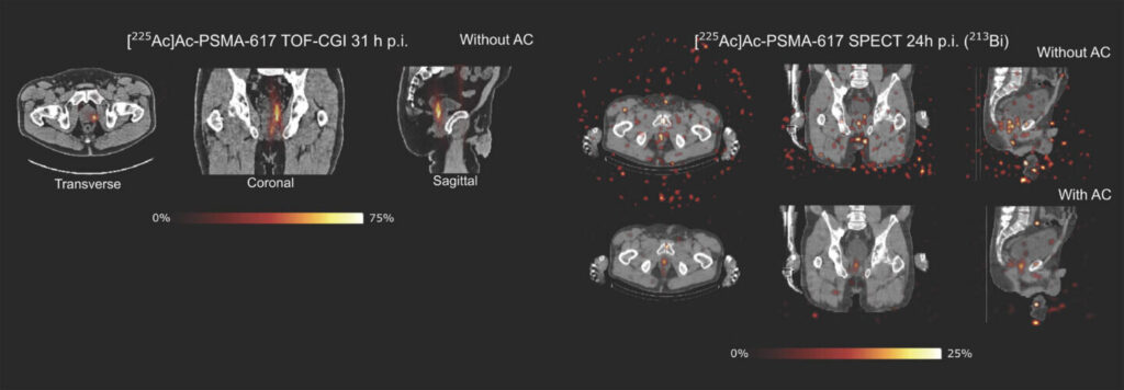
Caravaca and co-authors at UC San Francisco then conducted a pilot study with a patient who was previously diagnosed with prostate cancer through standard PET scans. A validation test confirmed that TOF-CGI in a PET scanner identified the same prostate tumors that were detected by the initial PET diagnostic scans. Subsequent experiments showed that TOF-CGI in PET detected more actinium-225 decays in the patient than SPECT, resulting in clearer images. The results represent the first TOF-CGI imaging of a human subject.
“This is an exciting first step. Clinicians want to know whether a radioisotope is doing its job. Did it stop the cancer cells from spreading? Or did new tumors emerge? Targeted alpha therapy imaging could help us answer those questions and more, but we still need an imaging modality to match. We think that our TOF-CGI technique could one day help us get there,” Caravaca said.
In future studies, Caravaca and his team hope to expand the new technique to other radioisotopes and test its efficiency with a full-body PET scanner.
Researchers from Berkeley Lab, UC San Francisco and Siemens Medical Solutions U.S. Inc. contributed to the study.
From particle physics to PET medical imaging: How Berkeley Lab became the birthplace of nuclear medicine
When Berkeley Lab founder Ernest Lawrence invented the cyclotron, the breakthrough not only revolutionized particle physics—it led to the birth of nuclear medicine. In 1935, Ernest Lawrence invited his brother John Lawrence, a physician, to join the lab and explore the use of cyclotron-produced radioisotopes for research in medicine and biology. The next year, John became the first to treat a cancer patient with a radioisotope that was produced in one of Ernest Lawrence’s cyclotrons.
More breakthroughs followed, and many of the radioisotopes used as biological tracers in research and medicine today, such as iodine-131, technetium-99m, carbon-14 and many others, were discovered at Berkeley Lab.
Berkeley Lab research has also led to advances in medical imaging. In the 1950s, Hal Anger developed the scintillation camera to image gamma rays emitted by radioisotopes. The scintillation camera—which later became known as the Anger camera—laid the foundation for today’s PET and SPECT scanners.
In the following decades, medical imaging has continued to evolve with innovations in detectors, computational techniques and electronics, with Berkeley Lab’s contributions to the first total-body PET scanner in 2015 representing a significant milestone that laid the groundwork for lower-dose, higher-resolution medical imaging. In more recent advancements, Berkeley Lab researchers are applying PET to understand the role of amyloid and tau proteins in Alzheimer’s disease.
Publication details
Javier Caravaca et al, Clinical Imaging of 225 Ac in a PET Scanner via Time-of-Flight Cascade γ-Ray Imaging, Journal of Nuclear Medicine (2026). DOI: 10.2967/jnumed.126.272594
Journal information:
Journal of Nuclear Medicine


Citation:
PET scans reveal alpha cancer therapy isotope with new algorithm in first human test (2026, October 1)
retrieved 1 October 2026
from https://medicalxpress.com/news/2026-10-pet-scans-reveal-alpha-cancer.html
This document is subject to copyright. Apart from any fair dealing for the purpose of private study or research, no
part may be reproduced without the written permission. The content is provided for information purposes only.

Khamrah by Lattafa for Men - 3.4 oz EDP Spray
4% Off
Ghost Sweetheart Eau de Toilette | Pineapple, Jasmine and Sandalwood | Perfume for Women 50 ml
50% Off
Marc Jacobs Dot Eau De Parfum for Women, 100 ml
42% Off
Ted Baker W Eau de Toilette for Her, Fig Leaf, White Peony and Violet Top Notes, Pink Orchid and Raspberry Middle Notes, 75ml
£11.77 (£15.69 / 100 ml) (as of 01/10/2026 05:09 GMT +01:00 - More infoProduct prices and availability are accurate as of the date/time indicated and are subject to change. Any price and availability information displayed on [relevant Amazon Site(s), as applicable] at the time of purchase will apply to the purchase of this product.)
Ted Baker Woman Pink Eau de Toilette Spray Floral Green Feminine Fragrance, Opening Notes are Fresh Peach, Bergamot and Tangerine with Warm Musk, Vanilla and Vetiver Base, 100ml
11% Off
Vera Wang Princess Eau de Toilette - 30 ml

Choco Musk 50ml Eau De Parfum for men and women | Chocolate Musk by Jannat Aromas
17% Off
Christina Aguilera Signature Eau de Parfum (50ml) Floral, Fruity & Exotic Scent, Luxury Fragrance for Women
9% Off
Calvin Klein - Eau De Toilette CKIN2U - Calvin Klein Women, Ladies Perfume, Women's Perfume, Calvin Klein Perfume, Calvin Klein One - 150 ml
5% Off
Jimmy Choo Flash Eau de Parfum, 60 ml (Pack of 1)
3% Off
Fruit of the Loom Men's Heavy T Shirt, White, XL UK
28% Off
ATNKE LED Lighted Beanie Cap,USB Rechargeable Running Hat Ultra Bright 4 LED Waterproof Light Winter Warm Gifts for Men and Women/Pink
17% Off
Men's 1/4 Zip Pullover UK Sale Clearance, Fleece Sweatshirt Casual Jumper Long Sleeve T-shirt Top Stand Collar Sweater Plain Pullover Sports Leisure Workwear Quarter Zip Sweater Lightweight Jumpers
£5.88 (as of 12/11/2025 00:52 GMT +01:00 - More infoProduct prices and availability are accurate as of the date/time indicated and are subject to change. Any price and availability information displayed on [relevant Amazon Site(s), as applicable] at the time of purchase will apply to the purchase of this product.)
Crevice Cleaning Brush, Bathroom Tile Groove Gap Cleaning Brush,Premium Crevice Cleaning Tool Aluminum Support with 15° Angle Magic Brush, Thin Brush for Home Kitchen
19% Off
Wireless Earbuds, Bluetooth 5.3 Headphones in Ear with HiFi Stereo Deep Bass, 4 ENC Noise Cancelling Mic Wireless Earphones 40H Playtime, Bluetooth Earbuds Dual LED Display, IP7 Waterproof, USB-C
42% Off