[
[

A cross-disciplinary research team has, for the first time, successfully applied NIR-II (1,000–3,000 nm) fluorescence video imaging during esophagectomy—a complex surgical procedure involving the resection of a diseased or cancerous portion of the esophagus, where the most dreaded complication, anastomotic leakage, a gastrointestinal defect at the suture line between the esophagus and the gastric conduit, affects 10–30% of patients and leads to high postoperative morbidity rate and even death.
The team’s findings have been published in Proceedings of the National Academy of Sciences.
In Hong Kong, about 30 patients have already benefited from esophagectomy performed with the aid of this new fluorescence imaging and video analysis technique, with highly promising outcomes to date.
The team was led by Professor Hongjie Dai (Sapientia Eminence Professor, Chair Professor in the Department of Chemistry, Department of Mechanical Engineering and School of Biomedical Sciences, Director of the Materials Institute of Life Sciences and Energy (MILES)) and Professor Simon Ying Kit Law (Chairperson and Cheung Kung-Hai Professor in Gastrointestinal Surgery, Chair Professor in Department of Surgery, the School of Clinical Medicine, HKUMed) at the University of Hong Kong (HKU).
By combining high-clarity NIR-II video imaging with rapid computational video analysis, the HKU research team’s pioneering technology enables operator-independent, objective delineation of boundaries between well-perfused and poorly perfused regions, providing precise guidance for resection and reconstruction to reduce the anastomotic leakage rate.
Challenges of current NIR-I fluorescence imaging in assessing gastric conduit perfusion during esophagectomy
Accurate, operator-independent assessment of tissue perfusion and selection of an optimal anastomotic site during esophagectomy are essential for surgeons to ensure the viability of the gastric conduit and to minimize the risk of anastomotic leakage.
Currently, near-infrared (NIR) fluorescence imaging using conventional dyes, such as indocyanine green (ICG), with a commercially available near-infrared I (NIR-I, 700–900 nm) instrument is used for diagnostics and intraoperative navigation.
Despite its widespread use, NIR-I angiography has several limitations, such as superficial penetration depth, high background signal and lower resolution caused by light scattering and tissue autofluorescence.
Further, assessing perfusion based on the suboptimal images relies heavily on the surgeon’s experience and subjective image interpretation, making it difficult to precisely determine the demarcation line of blood perfusion in the gastric conduit, a process critical to the outcome of the surgery.
Intraoperative NIR-II fluorescence-guided esophagectomy in the operating room
This study represents the first clinical use of NIR-II (1,000–3,000 nm) fluorescence imaging to visualize blood perfusion in the gastric conduits, affording markedly superior contrast and resolution compared to traditional NIR-I imaging.
NIR-II imaging was pioneered by Dai’s team in 2009 with mice, taking advantage of the reduced light scattering and tissue autofluorescence at light wavelengths above 1,000 nm. This breakthrough marks the first successful translation of NIR-II imaging into clinical practice for upper gastrointestinal surgery in human patients.
Highlights of the study include:
- The building of an imaging system that captures photographic images and NIR-II fluorescence images at the same time
- Much clearer blood flow images in NIR-II than traditional NIR-I, allowing more precise assessment of blood perfusion
- The combined use of a rapid intraoperative computational analysis of high-resolution blood perfusion NIR-II videos that identifies well-perfused and poorly perfused regions of the gastric conduits within one minute of dye administration.
Professor Hongjie Dai commented, “This is a great example of cross-disciplinary collaboration for translation of our imaging technique from mouse to man.”
Professor Simon Law added, “This innovation offers objective guidance for surgical decisions in esophagectomy. It enables surgeons to accurately assess blood supply, and remove the poorly perfused portion in order to perform the anastomosis at a well perfused area, thereby reducing the risk of anastomotic leakage. This not only enhances surgical safety but also represents a significant breakthrough in esophagectomy.”
This study is part of Hong Kong’s Research, Academic and Industry Sectors One-plus Scheme (RAISe+), the JC STEM Lab of Nanoscience and Nanomedicine and Materials Institute of Life Sciences and Energy (MILES) in Shenzhen. This RAISe+ project is revolutionizing surgery with advanced dyes and imaging tools that work in the NIR-II window.
This technology gives surgeons “infrared vision” to see tumors, blood vessels, and organs clearly and deeply during operations.
The team’s solution helps overcome current limitations of shallow imaging depth and low image quality, enabling precise tumor removal, ureter detection, and sentinel lymph node identification, making surgeries safer and more effective for patients.
More information:
Feifei Wang et al, Guiding esophagectomy with intraoperative NIR-II fluorescence video imaging and rapid computation, Proceedings of the National Academy of Sciences (2025). DOI: 10.1073/pnas.2517785122
Citation:
For the first time, next-gen infrared fluorescence imaging helps surgeons see blood perfusion during esophageal surgery (2025, December 9)
retrieved 9 December 2025
from https://medicalxpress.com/news/2025-12-gen-infrared-fluorescence-imaging-surgeons.html
This document is subject to copyright. Apart from any fair dealing for the purpose of private study or research, no
part may be reproduced without the written permission. The content is provided for information purposes only.

Khamrah by Lattafa for Men - 3.4 oz EDP Spray
4% Off
Ghost Sweetheart Eau de Toilette | Pineapple, Jasmine and Sandalwood | Perfume for Women 50 ml
50% Off
Marc Jacobs Dot Eau De Parfum for Women, 100 ml
42% Off
Ted Baker W Eau de Toilette for Her, Fig Leaf, White Peony and Violet Top Notes, Pink Orchid and Raspberry Middle Notes, 75ml
£11.77 (£15.69 / 100 ml) (as of 30/05/2026 03:24 GMT +01:00 - More infoProduct prices and availability are accurate as of the date/time indicated and are subject to change. Any price and availability information displayed on [relevant Amazon Site(s), as applicable] at the time of purchase will apply to the purchase of this product.)
Ted Baker Woman Pink Eau de Toilette Spray Floral Green Feminine Fragrance, Opening Notes are Fresh Peach, Bergamot and Tangerine with Warm Musk, Vanilla and Vetiver Base, 100ml
11% Off
Vera Wang Princess Eau de Toilette - 30 ml
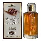
Choco Musk 50ml Eau De Parfum for men and women | Chocolate Musk by Jannat Aromas
17% Off
Christina Aguilera Signature Eau de Parfum (50ml) Floral, Fruity & Exotic Scent, Luxury Fragrance for Women
9% Off
Calvin Klein - Eau De Toilette CKIN2U - Calvin Klein Women, Ladies Perfume, Women's Perfume, Calvin Klein Perfume, Calvin Klein One - 150 ml
5% Off
Jimmy Choo Flash Eau de Parfum, 60 ml (Pack of 1)
3% Off
Fruit of the Loom Men's Heavy T Shirt, White, XL UK
28% Off
ATNKE LED Lighted Beanie Cap,USB Rechargeable Running Hat Ultra Bright 4 LED Waterproof Light Winter Warm Gifts for Men and Women/Pink
17% Off
Men's 1/4 Zip Pullover UK Sale Clearance, Fleece Sweatshirt Casual Jumper Long Sleeve T-shirt Top Stand Collar Sweater Plain Pullover Sports Leisure Workwear Quarter Zip Sweater Lightweight Jumpers
£5.88 (as of 12/11/2025 00:52 GMT +01:00 - More infoProduct prices and availability are accurate as of the date/time indicated and are subject to change. Any price and availability information displayed on [relevant Amazon Site(s), as applicable] at the time of purchase will apply to the purchase of this product.)
Crevice Cleaning Brush, Bathroom Tile Groove Gap Cleaning Brush,Premium Crevice Cleaning Tool Aluminum Support with 15° Angle Magic Brush, Thin Brush for Home Kitchen
19% Off
Wireless Earbuds, Bluetooth 5.3 Headphones in Ear with HiFi Stereo Deep Bass, 4 ENC Noise Cancelling Mic Wireless Earphones 40H Playtime, Bluetooth Earbuds Dual LED Display, IP7 Waterproof, USB-C
42% Off