 [
[

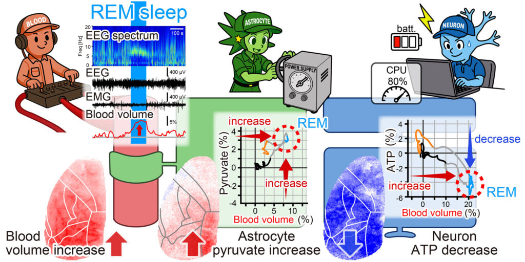
REM sleep is already called “paradoxical sleep.” The body is largely still, yet the brain is highly active. In humans, this is also the stage most closely associated with vivid dreaming. Our study, published in Communications Biology, has revealed another paradox hidden within REM sleep: Brain blood volume increased, but the ATP available inside neurons decreased.
We began with a simple question: How does the sleeping brain adjust its energy supply to match changing patterns of information processing?
The brain is an energy-hungry organ. Neurons need ATP to generate electrical signals, restore ion gradients and modify synapses. Blood vessels deliver oxygen and glucose, while astrocytes—glial cells that contact both vessels and neurons—help process and distribute metabolic substrates.
To understand this system, we followed three different signals: brain blood volume, astrocytic pyruvate and neuronal ATP.
This was technically challenging. Opening the skull can disturb intracranial pressure, vascular activity and glial function. We therefore observed the cerebral cortex through the native, unthinned skull of living mice. After exposing the skull, we coated it with a transparent UV-curable resin and used wide-field fluorescence imaging while the animals naturally moved between non-REM sleep, REM sleep and wakefulness.
The sleeping brain continuously adjusts its vascular support
During non-REM sleep, we found that fluctuations in theta-band brain activity predicted changes in brain blood volume approximately four to five seconds later. This was surprising because non-REM sleep is more commonly associated with strong delta activity. The weaker theta fluctuations, however, closely tracked the subsequent vascular response.
To us, this suggested that blood-volume changes during sleep are not simply passive background fluctuations. Even in a sleeping brain, vascular support appears to be continuously adjusted to ongoing neuronal activity.
We also found a spatial pattern in these fluctuations. During non-REM sleep, relatively fast blood-volume waves repeatedly propagated from anterior to posterior cortical regions, crossing much of the observed cortex in approximately one second.

The brain prepares for REM sleep before REM is detected
The transition into REM sleep was even more striking. Brain blood volume began to rise about 50 seconds before REM sleep could be conventionally identified from brain and muscle recordings. The increase began in the posterior cortex and then spread toward anterior regions over approximately 15 to 20 seconds.
In other words, the brain appeared to begin preparing for REM sleep before the usual electrical signs of REM were fully established.
At that point, we expected intracellular energy signals to rise together with the vascular signal. This expectation seemed reasonable. When we pharmacologically dilated cerebral vessels, both astrocytic pyruvate and neuronal ATP increased.
Natural REM sleep did not follow that pattern.
Our initial expectation was wrong
Astrocytic pyruvate increased during REM sleep, particularly in posterior cortical regions. Neuronal ATP, however, moved in the opposite direction.

That was the result that changed the direction of our study. We were no longer looking only at sleep-related vascular dynamics. We had uncovered a dissociation between vascular delivery, astrocytic metabolism and neuronal energy availability.
The key results can be stated simply:
- Brain blood volume increased.
- Astrocytic pyruvate increased.
- Neuronal ATP decreased.
Why would the brain allow neuronal ATP to fall during a state of intense internal processing?
We do not yet know. One possibility is that neurons consume ATP rapidly during REM-specific processes, including synaptic reorganization and communication between the hippocampus and cortex.
Another possibility is that the transfer of metabolic substrates from astrocytes to neurons changes during REM sleep. A third possibility is that mitochondrial ATP production becomes temporarily less efficient.
These mechanisms are not mutually exclusive. REM sleep may combine increased ATP consumption with altered substrate transfer and altered energy production.

What vascular imaging can—and cannot—tell us
Our findings also made us think differently about hemodynamic brain imaging.
Functional MRI does not measure neuronal activity directly. It infers brain activity from changes in blood flow, blood volume and blood oxygenation.
These vascular signals are extremely useful, but our study shows that they do not tell the whole metabolic story. During REM sleep, the vascular signal increased while neuronal ATP moved in the opposite direction.
We therefore think of brain energy regulation as a multilayered process. Blood vessels adjust the delivery of oxygen and glucose. Astrocytes process, store and redistribute metabolic substrates. Neurons use ATP to perform information processing.
The biological brain may achieve its extraordinary energy efficiency not by supplying every cell uniformly, but by reallocating limited resources according to its current computational state.
REM sleep offers a natural example of this strategy. More blood did not simply mean more neuronal energy. Instead, the relationship among vessels, astrocytes and neurons was reorganized.
Have you ever awakened from a vivid dream feeling strangely tired? Our experiment did not test human fatigue, so any connection remains speculative. But the cellular phenomenon itself is clear: During REM sleep, neuronal ATP can fall even while brain blood volume rises.
Understanding why this happens may help us uncover not only the biology of sleep and memory, but also the principles that allow biological intelligence to operate with such remarkable energy efficiency.
This story is part of Science X Dialog, where researchers can report findings from their published research articles. Visit this page for information about Science X Dialog and how to participate.
Publication details
Yusuke Takahashi et al, Energy paradox in REM sleep: balancing supply and consumption in brain metabolism, Communications Biology (2026). DOI: 10.1038/s42003-026-10646-6
Journal information:
Communications Biology
Key medical concepts


Ko Matsui is a professor of Super-network Brain Physiology at Tohoku University, Japan. His laboratory investigates how neurons, astrocytes, blood vessels and whole-body signals interact to support brain information processing, learning, sleep and metabolism. His research focuses on the dynamic coupling between neural computation and metabolic energy allocation in the living brain.
Citation:
When more blood does not mean more neuronal energy during REM sleep (2026, August 2)
retrieved 2 August 2026
from https://medicalxpress.com/news/2026-07-blood-neuronal-energy-rem.html
This document is subject to copyright. Apart from any fair dealing for the purpose of private study or research, no
part may be reproduced without the written permission. The content is provided for information purposes only.

Khamrah by Lattafa for Men - 3.4 oz EDP Spray
4% Off
Ghost Sweetheart Eau de Toilette | Pineapple, Jasmine and Sandalwood | Perfume for Women 50 ml
50% Off
Marc Jacobs Dot Eau De Parfum for Women, 100 ml
42% Off
Ted Baker W Eau de Toilette for Her, Fig Leaf, White Peony and Violet Top Notes, Pink Orchid and Raspberry Middle Notes, 75ml
£11.77 (£15.69 / 100 ml) (as of 03/08/2026 04:05 GMT +01:00 - More infoProduct prices and availability are accurate as of the date/time indicated and are subject to change. Any price and availability information displayed on [relevant Amazon Site(s), as applicable] at the time of purchase will apply to the purchase of this product.)
Ted Baker Woman Pink Eau de Toilette Spray Floral Green Feminine Fragrance, Opening Notes are Fresh Peach, Bergamot and Tangerine with Warm Musk, Vanilla and Vetiver Base, 100ml
11% Off
Vera Wang Princess Eau de Toilette - 30 ml

Choco Musk 50ml Eau De Parfum for men and women | Chocolate Musk by Jannat Aromas
17% Off
Christina Aguilera Signature Eau de Parfum (50ml) Floral, Fruity & Exotic Scent, Luxury Fragrance for Women
9% Off
Calvin Klein - Eau De Toilette CKIN2U - Calvin Klein Women, Ladies Perfume, Women's Perfume, Calvin Klein Perfume, Calvin Klein One - 150 ml
5% Off
Jimmy Choo Flash Eau de Parfum, 60 ml (Pack of 1)
3% Off
Fruit of the Loom Men's Heavy T Shirt, White, XL UK
28% Off
ATNKE LED Lighted Beanie Cap,USB Rechargeable Running Hat Ultra Bright 4 LED Waterproof Light Winter Warm Gifts for Men and Women/Pink
17% Off
Men's 1/4 Zip Pullover UK Sale Clearance, Fleece Sweatshirt Casual Jumper Long Sleeve T-shirt Top Stand Collar Sweater Plain Pullover Sports Leisure Workwear Quarter Zip Sweater Lightweight Jumpers
£5.88 (as of 12/11/2025 00:52 GMT +01:00 - More infoProduct prices and availability are accurate as of the date/time indicated and are subject to change. Any price and availability information displayed on [relevant Amazon Site(s), as applicable] at the time of purchase will apply to the purchase of this product.)
Crevice Cleaning Brush, Bathroom Tile Groove Gap Cleaning Brush,Premium Crevice Cleaning Tool Aluminum Support with 15° Angle Magic Brush, Thin Brush for Home Kitchen
19% Off
Wireless Earbuds, Bluetooth 5.3 Headphones in Ear with HiFi Stereo Deep Bass, 4 ENC Noise Cancelling Mic Wireless Earphones 40H Playtime, Bluetooth Earbuds Dual LED Display, IP7 Waterproof, USB-C
42% Off