
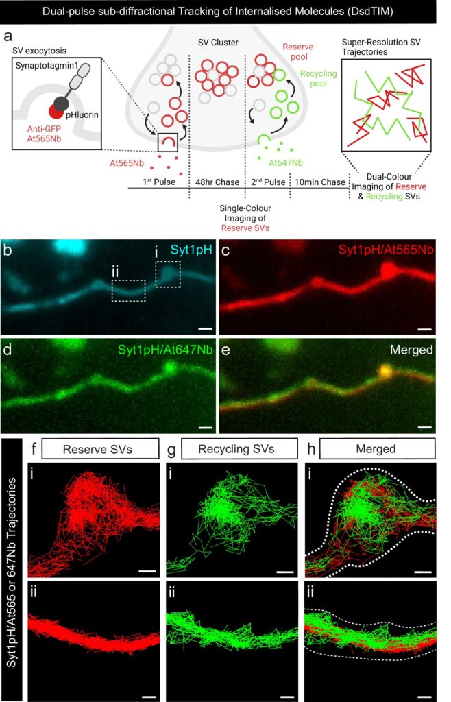
 protocol. b Representative epifluorescence image of an axonal segment expressing Syt1pH acquired before incubation with At647Nb. The dashed boxes in (b) highlight (i) a presynaptic compartment and (ii) a peri-synaptic axonal segment. c–e Maximum intensity projection of (c) Syt1pH/At565Nb, (d) Syt1pH/At647Nb and (e) merged maximum intensity projection. f Trajectory map of tracked reserve SVs containing Syt1pH bound by At565Nb in the (i) presynapse and (ii) axonal segment. g Trajectory map of tracked recycling SVs containing Syt1pH bound by At647Nb in the (i) presynapse and (ii) axonal segment. h Merged trajectory maps. Credit: Nature Communications (2024). DOI: 10.1038/s41467-024-46256-1")
Researchers from UQ’s Queensland Brain Institute (QBI) have revealed the pivotal role played by Synapsin 2a proteins in orchestrating the organization and mobility of synaptic vesicles within live neurons.
Shanley Longfield, a Ph.D. student in the Meunier lab, explained that she used super resolution microscopy, simultaneously tracking key proteins and synaptic vesicles, to make this discovery.
This study was a collaboration with the Ecole Normale Superieure (France), the University of Cambridge (UK), and the Temasek Lifesciences Laboratory (Singapore). The article was published in Nature Communications.
“We used a novel technique, known as dual-pulse sub-diffractional tracking of internalized molecules, developed in our lab, to peer into the inner workings of live hippocampal neurons,” Longfield said.
“One of our main findings revolves around the role of Synapsin 2a tetramerization in controlling the clustering and mobility of reserve synaptic vesicles.
“The reserve synaptic vesicles, which constitute a crucial backup reservoir of neurotransmitters, display lower mobility and are less responsive to high-frequency stimulation compared to their recycling pool counterparts.
“This revelation underscores the intricate balance maintained within neurons to ensure efficient neurotransmitter release under varying physiological conditions.
“Our study also sheds light on the distinct mobility patterns of reserve and recycling synaptic vesicles at different neuronal terminals, emphasizing the nuanced regulation of these dynamics by Synapsin proteins.
“Synapsins serve as molecular tethers, anchoring the synaptic vesicles within clusters and fine-tuning their movements, ultimately influencing neurotransmitter release and neuronal communication.
“In earlier research, we have shown that tau, a protein involved in Alzheimer’s disease, controls the dynamic clustering of the important subpopulation of recycling vesicles at the presynapse critical for neuronal communication.
“We found that tau forms small and transient nanoscale biomolecular condensates that can selectively capture these vesicles at the presynapse.
“Now we have tracked the protein—Synapsin 2a—at the same time as the reserve vesicles and shown Synapsin 2a can form tetramers that cross-link synaptic vesicles at the presynapse.”
Professor Fred Meunier said the next step is to study how the two mechanisms, one involving tau and the other Synapsin 2a, “talk to each other” to drive neuronal communication.
“Understanding how condensates and the cross-linking mechanisms coordinate the clustering of synaptic vesicles to allow the presynapse to communicate will be a challenging but exciting endeavor,” Meunier said.
“The implications of this research extend far beyond the realm of basic neuroscience.
“Understanding the precise mechanisms governing synaptic vesicle dynamics deepens our knowledge of fundamental brain processes and holds potential therapeutic applications for neurological disorders, such as Alzheimer’s disease, characterized by disruptions in synaptic function.”
More information:
Shanley F. Longfield et al, Synapsin 2a tetramerisation selectively controls the presynaptic nanoscale organisation of reserve synaptic vesicles, Nature Communications (2024). DOI: 10.1038/s41467-024-46256-1
Provided by
Queensland Brain Institute
Citation:
Unraveling the mysteries of the presynapse with super resolution microscopy (2024, March 20)
retrieved 20 March 2024
from https://medicalxpress.com/news/2024-03-unraveling-mysteries-presynapse-super-resolution.html
This document is subject to copyright. Apart from any fair dealing for the purpose of private study or research, no
part may be reproduced without the written permission. The content is provided for information purposes only.

Khamrah by Lattafa for Men - 3.4 oz EDP Spray
4% Off
Ghost Sweetheart Eau de Toilette | Pineapple, Jasmine and Sandalwood | Perfume for Women 50 ml
50% Off
Marc Jacobs Dot Eau De Parfum for Women, 100 ml
42% Off
Ted Baker W Eau de Toilette for Her, Fig Leaf, White Peony and Violet Top Notes, Pink Orchid and Raspberry Middle Notes, 75ml
£11.77 (£15.69 / 100 ml) (as of 12/07/2026 03:56 GMT +01:00 - More infoProduct prices and availability are accurate as of the date/time indicated and are subject to change. Any price and availability information displayed on [relevant Amazon Site(s), as applicable] at the time of purchase will apply to the purchase of this product.)
Ted Baker Woman Pink Eau de Toilette Spray Floral Green Feminine Fragrance, Opening Notes are Fresh Peach, Bergamot and Tangerine with Warm Musk, Vanilla and Vetiver Base, 100ml
11% Off
Vera Wang Princess Eau de Toilette - 30 ml

Choco Musk 50ml Eau De Parfum for men and women | Chocolate Musk by Jannat Aromas
17% Off
Christina Aguilera Signature Eau de Parfum (50ml) Floral, Fruity & Exotic Scent, Luxury Fragrance for Women
9% Off
Calvin Klein - Eau De Toilette CKIN2U - Calvin Klein Women, Ladies Perfume, Women's Perfume, Calvin Klein Perfume, Calvin Klein One - 150 ml
5% Off
Jimmy Choo Flash Eau de Parfum, 60 ml (Pack of 1)
3% Off
Fruit of the Loom Men's Heavy T Shirt, White, XL UK
28% Off
ATNKE LED Lighted Beanie Cap,USB Rechargeable Running Hat Ultra Bright 4 LED Waterproof Light Winter Warm Gifts for Men and Women/Pink
17% Off
Men's 1/4 Zip Pullover UK Sale Clearance, Fleece Sweatshirt Casual Jumper Long Sleeve T-shirt Top Stand Collar Sweater Plain Pullover Sports Leisure Workwear Quarter Zip Sweater Lightweight Jumpers
£5.88 (as of 12/11/2025 00:52 GMT +01:00 - More infoProduct prices and availability are accurate as of the date/time indicated and are subject to change. Any price and availability information displayed on [relevant Amazon Site(s), as applicable] at the time of purchase will apply to the purchase of this product.)
Crevice Cleaning Brush, Bathroom Tile Groove Gap Cleaning Brush,Premium Crevice Cleaning Tool Aluminum Support with 15° Angle Magic Brush, Thin Brush for Home Kitchen
19% Off
Wireless Earbuds, Bluetooth 5.3 Headphones in Ear with HiFi Stereo Deep Bass, 4 ENC Noise Cancelling Mic Wireless Earphones 40H Playtime, Bluetooth Earbuds Dual LED Display, IP7 Waterproof, USB-C
42% Off


