 [


Researchers at The Hong Kong Polytechnic University (PolyU) have developed an imaging biomarker, the Tumour Heterogeneity Index (THI), which enables noninvasive assessment of breast cancer patients’ response to AKT inhibitors before treatment, helping clinicians identify patients most likely to benefit from therapy. The study found that patients with a high THI achieved an estimated pathological complete response (pCR) rate of 56.1%, significantly higher than that of patients with a low THI. The technology could help patients receive the most appropriate treatment at an earlier stage, reduce unnecessary side effects and enable clinicians to better tailor treatment strategies to individual patients.
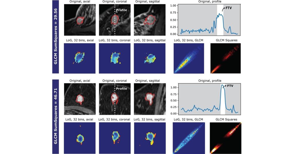
Cai Jing, head of the Department of Health Technology and Informatics (HTI) and chair professor of medical physics and intelligent oncology at PolyU, together with Dr. Teng Xinzhi, research assistant professor of HTI, and their research team, applied advanced radiomics techniques to develop THI using data from dynamic contrast-enhanced magnetic resonance imaging (DCE-MRI). DCE-MRI is widely used for cancer diagnosis and treatment planning. The team has further extended its application to the assessment of drug response, enabling clinicians to noninvasively predict treatment outcomes before therapy begins.
The study focused on patients with high-risk, early-stage breast cancer and evaluated their response to the investigational AKT inhibitor MK2206 when added to neoadjuvant chemotherapy before surgery. The AKT/mTOR/PI3K signaling pathway plays an important role in promoting tumor growth and survival and has therefore long been regarded as an important therapeutic target in cancer treatment.
Although the AKT inhibitor MK2206 has previously been shown to improve pCR in the large-scale I-SPY2 clinical trial, its overall efficacy remained limited, with marked variations in treatment response among aggressive subtypes including HER2-negative, hormone receptor-negative (HR−) and triple-negative breast cancer. Identifying patients most likely to benefit before treatment has therefore remained an important clinical challenge.
Cai said, “This challenge highlights the importance of accurately predicting treatment response before therapy begins. Our study presents an effective strategy for identifying patients who are most likely to benefit from treatment, with the potential to enhance the clinical value of drugs that may not have achieved the expected outcomes in large-scale clinical trials. THI is more than an imaging analysis tool. It quantifies the complexity of a tumor’s internal structure, transforming assessment that has traditionally relied on experience into an objective, data-driven approach.”
The findings showed that THI outperformed conventional immunohistochemistry (IHC)-based receptor testing in identifying patients suitable for AKT inhibitor treatment and in characterizing the tumor microenvironment. By analyzing the immune cells and vascular environment surrounding the tumor, clinicians could more accurately assess drug response and the risk of treatment resistance, thereby informing more effective treatment strategies.
The study found that patients with a high THI had an estimated pCR rate of up to 56.1%, significantly higher than that of patients with a low THI. Promising results were also observed among patients with triple-negative breast cancer, a subtype that is typically more difficult to treat. The study further demonstrated that THI was independently and specifically associated with response to AKT inhibition, underscoring its value as a predictive biomarker. The findings have been published in the journal Breast Cancer Research.
Cai said, “Through THI, we have the opportunity to identify, before treatment begins, patients who are most likely to benefit, while sparing others from treatments with limited effectiveness. This can not only improve overall treatment outcomes but also reduce unnecessary side effects, bringing us an important step closer to truly personalized cancer treatment. By using noninvasive imaging examinations, the approach could also reduce reliance on costly genetic sequencing in the future, making precision medicine more widely applicable in routine clinical practice.”
Compared with conventional biopsies, which can analyze only a localized sample of tumor tissue, THI enables comprehensive assessment of the entire tumor, reducing the risk of sampling errors arising from differences in biopsy location while also minimizing the discomfort associated with repeated biopsies. The research team has also conducted rigorous repeatability and reproducibility validation, demonstrating that the technology can maintain stable and reliable performance across different imaging equipment and health care institutions, highlighting its potential for broad clinical application.
Teng said, “DCE-MRI is already widely used in breast cancer diagnosis, providing a rich source of readily available clinical data for our research. Using advanced radiomics algorithms, we transform imaging information originally used for diagnosis into a tool for treatment decision-making, quantifying tumor heterogeneity and developing a predictive scoring system.”
Building on its analysis of tumor vascular texture features, the research team has also developed a new model, DCE-MRI Vascularisation-based Response Tracking (DCE-VASC-TRACK), which uses longitudinal DCE-MRI to noninvasively monitor changes in tumor vasculature. The model integrates repeatable vascular texture features associated with pCR at surgery with hormone receptor status, age, baseline functional tumor volume (FTV) and mid-treatment changes in FTV. Compared with the use of clinical factors and FTV measurements alone, the model significantly improves the prediction of pCR.
This related study has been published in Radiology: Artificial Intelligence. The findings showed that, compared with assessing tumor size alone, DCE-VASC-TRACK can more effectively monitor response to neoadjuvant chemotherapy in patients with HER2-negative breast cancer. By capturing changes in the tumor microenvironment, the model could serve as a practical and cost-effective alternative for predicting treatment response while reducing reliance on invasive multi-omics testing.
The research team noted that different cancers have distinct biological characteristics and clinical needs. For example, breast cancer treatment places particular emphasis on precisely matching patients with appropriate drugs, whereas treatment for nasopharyngeal carcinoma focuses more on reducing treatment toxicity while maintaining efficacy. The team will therefore tailor the application of technology to the clinical needs of different cancer types. The researchers believe that this approach could in the future be extended to a wider range of cancers and treatment strategies, further advancing the development of precision medicine.
Publication details
Xinzhi Teng et al., Tumor heterogeneity index from DCE-MRI for AKT-inhibition responder identification and reveals hot immune microenvironment for patients with breast cancer: a multi-omics analysis of I-SPY2 trial, Breast Cancer Research (2026). DOI: 10.1186/s13058-026-02219-6
Xinzhi Teng et al., Longitudinal DCE MRI Vascular Textures: Radiologic and Biologic Insights for pCR Prediction in HER2-Negative Breast Cancer, Radiology: Artificial Intelligence (2026). DOI: 10.1148/ryai.250734
Journal information:
Breast Cancer Research
Clinical categories


Citation:
Tumor index can identify breast cancer patients’ drug response (2026, November 1)
retrieved 1 November 2026
from https://medicalxpress.com/news/2026-10-tumor-index-breast-cancer-patients.html
This document is subject to copyright. Apart from any fair dealing for the purpose of private study or research, no
part may be reproduced without the written permission. The content is provided for information purposes only.

Khamrah by Lattafa for Men - 3.4 oz EDP Spray
4% Off
Ghost Sweetheart Eau de Toilette | Pineapple, Jasmine and Sandalwood | Perfume for Women 50 ml
50% Off
Marc Jacobs Dot Eau De Parfum for Women, 100 ml
42% Off
Ted Baker W Eau de Toilette for Her, Fig Leaf, White Peony and Violet Top Notes, Pink Orchid and Raspberry Middle Notes, 75ml
£11.77 (£15.69 / 100 ml) (as of 02/11/2026 05:39 GMT +00:00 - More infoProduct prices and availability are accurate as of the date/time indicated and are subject to change. Any price and availability information displayed on [relevant Amazon Site(s), as applicable] at the time of purchase will apply to the purchase of this product.)
Ted Baker Woman Pink Eau de Toilette Spray Floral Green Feminine Fragrance, Opening Notes are Fresh Peach, Bergamot and Tangerine with Warm Musk, Vanilla and Vetiver Base, 100ml
11% Off
Vera Wang Princess Eau de Toilette - 30 ml

Choco Musk 50ml Eau De Parfum for men and women | Chocolate Musk by Jannat Aromas
17% Off
Christina Aguilera Signature Eau de Parfum (50ml) Floral, Fruity & Exotic Scent, Luxury Fragrance for Women
9% Off
Calvin Klein - Eau De Toilette CKIN2U - Calvin Klein Women, Ladies Perfume, Women's Perfume, Calvin Klein Perfume, Calvin Klein One - 150 ml
5% Off
Jimmy Choo Flash Eau de Parfum, 60 ml (Pack of 1)
3% Off
Fruit of the Loom Men's Heavy T Shirt, White, XL UK
28% Off
ATNKE LED Lighted Beanie Cap,USB Rechargeable Running Hat Ultra Bright 4 LED Waterproof Light Winter Warm Gifts for Men and Women/Pink
17% Off
Men's 1/4 Zip Pullover UK Sale Clearance, Fleece Sweatshirt Casual Jumper Long Sleeve T-shirt Top Stand Collar Sweater Plain Pullover Sports Leisure Workwear Quarter Zip Sweater Lightweight Jumpers
£5.88 (as of 11/11/2025 23:52 GMT +00:00 - More infoProduct prices and availability are accurate as of the date/time indicated and are subject to change. Any price and availability information displayed on [relevant Amazon Site(s), as applicable] at the time of purchase will apply to the purchase of this product.)
Crevice Cleaning Brush, Bathroom Tile Groove Gap Cleaning Brush,Premium Crevice Cleaning Tool Aluminum Support with 15° Angle Magic Brush, Thin Brush for Home Kitchen
19% Off
Wireless Earbuds, Bluetooth 5.3 Headphones in Ear with HiFi Stereo Deep Bass, 4 ENC Noise Cancelling Mic Wireless Earphones 40H Playtime, Bluetooth Earbuds Dual LED Display, IP7 Waterproof, USB-C
42% Off