

Bioengineers and neuroscientists at the Wu Tsai Neurosciences Institute at Stanford University have developed a highly sensitive tool for detecting brain cells’ subtlest electrical signals.
Published in the November 2024 issue of Neuron, this breakthrough allows scientists, for the first time, to visualize the full spectrum of electrical communication between human brain cells in real time. This dramatically enhances researchers’ ability to understand brain signaling and the abnormalities linked to disorders such as autism and epilepsy.
Every sensation, memory, and thought we experience is encoded in the flickering signals that ceaselessly course through our brain’s vast neural networks. Neurons “hear” these electrical whispers, their broad-branching dendrites intercepting messages through synaptic connections from thousands of informants.
Each message, passed encoded in a packet of chemical neurotransmitters, triggers a tiny electrical response at the synapse—a few thousandths of a volt or less, fading away within milliseconds. Most of these are barely perceptible by the cell.
But dozens of overlapping messages can add up to something meaningful, triggering a roughly 100 millivolt “spike” or “action potential” that the cell passes along to all its contacts in the network, distributing its own cascade of chemical messages, and continuing the electrical flux that, for example, allows you to read this sentence.
A challenge for neuroscientists since the first half of the 20th century has been how to detect these tiny signals, to decode the electrical language of the brain. Neuronal spikes, and eventually the tiny “synaptic potentials,” were first detected using ingenious advances in recording electrodes and preparations of isolated neurons and brain slices. But neuroscientists have long desired reliable ways to visualize the full spectrum of electrical flux of active brain circuits in real time—ideally in the human brain.
The new research makes this possible for the first time with a new genetically encoded voltage indicator called ASAP5, the latest in a family of cutting-edge voltage sensors developed by Stanford chemical engineer Michael Lin, an associate professor of neurobiology and bioengineering and member of the Wu Tsai Neurosciences Institute.
A new level of sensitivity for studying synaptic activity
The new advance builds on a foundational collaboration between Lin and computational biophysicist Ron Dror, the Cheriton Family Professor of Computer Science in the Stanford Artificial Intelligence Lab, which was catalyzed by a seed grant in 2017. That work was critical to developing ASAP5’s predecessors, ASAP3 and ASAP4.
These tools—and others developed by other laboratories—have become common means of observing electrical activity in cultured neurons and the brains of animals such as mice and flies. But these tools have so far been limited to detecting neuronal spikes and larger “subthreshold” synaptic signals. Getting to the level of sensitivity needed to detect even the quietest whispers of synaptic activity had remained a challenge.
“The unsolved problem was that voltage indicators haven’t had the sensitivity to detect individual synaptic events reliably in neurons, and haven’t been used to look at signaling between human neurons,” said Lin, the study’s senior author.
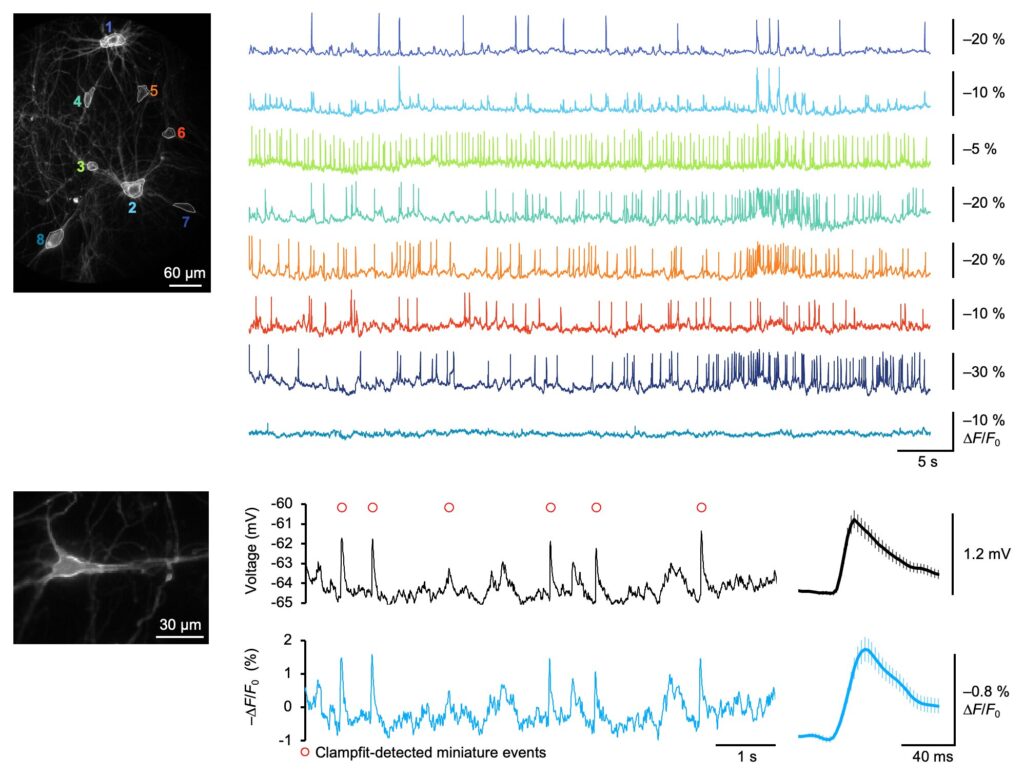
The new study found a solution. By screening thousands of genetic variants, the team engineered an ASAP molecule optimized to detect voltage changes specifically around the normal resting voltage of neurons. With the resulting sensor, researchers were able to detect even the random spontaneous release of individual packets of neurotransmitter—the smallest possible synaptic signal.
“Because of ASAP5’s improved sensitivity and speed, we can detect the small electrical impulses induced by the reception of single synaptic vesicles either locally at the synapse or at the cell body. This gave us the ability to observe how these impulses propagate from the synapse to the cell body,” Lin said.
Insights into neuronal signaling
These advancements not only address long-standing challenges in voltage detection but also enable new discoveries in how neurons communicate through these subtle electrical signals.
The tool has already allowed the research team to make new observations about how cells sum up the messages from their most far-flung informants. “Fascinatingly, we found that synapses that are farther away from the cell body initiate larger responses,” Lin explained.
In other words, neurons tune the most distant synapses on their extensive dendrites to be electrically “louder” so their input is not drowned out by synapses closer to the cell body.
Promise against neurological disease
ASAP5’s ability to detect synaptic potentials caused by the spontaneous release of single neurotransmitter packets—often referred to as synaptic “noise”—is not only an impressive marker of its sensitivity but may have important uses in studying neurological diseases.
Various disorders of the nervous system—from autism to epilepsy—involve changes in the basic properties of synapses, and so enabling scientists to observe the smallest whispers of synaptic signaling will be a valuable tool for scientists to understand, diagnose, and eventually treat these disorders.
The research team tested the tool across multiple common laboratory preparations and model organisms, including lab-grown human brain cells.
“We established that we could detect synaptic events in human neurons derived from human stem cells, and we could visualize coordination between multiple human neurons, opening up the possibility of rapidly assessing how mutations associated with disease affect the activity of synapses and the ability of neurons to fire within networks,” Lin said.
The work was done in collaboration with Tom Clandinin, Tom Südhof, Jun Ding and Marius Wernig at Stanford, and with Ed Boyden (MIT), Na Ji and Dan Feldman (Berkeley), Peyman Golshani (UCLA), and Stéphane Dieudonné (École Normale Supérieure). The two co-lead authors were Ph.D. student Alex Hao and postdoc Sungmoo Lee.
More information:
Yukun A. Hao et al, A fast and responsive voltage indicator with enhanced sensitivity for unitary synaptic events, Neuron (2024). DOI: 10.1016/j.neuron.2024.08.019
Citation:
New voltage indicator enables ultra-sensitive synaptic imaging (2024, October 30)
retrieved 30 October 2024
from https://medicalxpress.com/news/2024-10-voltage-indicator-enables-ultra-sensitive.html
This document is subject to copyright. Apart from any fair dealing for the purpose of private study or research, no
part may be reproduced without the written permission. The content is provided for information purposes only.

Khamrah by Lattafa for Men - 3.4 oz EDP Spray
4% Off
Ghost Sweetheart Eau de Toilette | Pineapple, Jasmine and Sandalwood | Perfume for Women 50 ml
50% Off
Marc Jacobs Dot Eau De Parfum for Women, 100 ml
42% Off
Ted Baker W Eau de Toilette for Her, Fig Leaf, White Peony and Violet Top Notes, Pink Orchid and Raspberry Middle Notes, 75ml
£11.77 (£15.69 / 100 ml) (as of 19/07/2026 04:00 GMT +01:00 - More infoProduct prices and availability are accurate as of the date/time indicated and are subject to change. Any price and availability information displayed on [relevant Amazon Site(s), as applicable] at the time of purchase will apply to the purchase of this product.)
Ted Baker Woman Pink Eau de Toilette Spray Floral Green Feminine Fragrance, Opening Notes are Fresh Peach, Bergamot and Tangerine with Warm Musk, Vanilla and Vetiver Base, 100ml
11% Off
Vera Wang Princess Eau de Toilette - 30 ml

Choco Musk 50ml Eau De Parfum for men and women | Chocolate Musk by Jannat Aromas
17% Off
Christina Aguilera Signature Eau de Parfum (50ml) Floral, Fruity & Exotic Scent, Luxury Fragrance for Women
9% Off
Calvin Klein - Eau De Toilette CKIN2U - Calvin Klein Women, Ladies Perfume, Women's Perfume, Calvin Klein Perfume, Calvin Klein One - 150 ml
5% Off
Jimmy Choo Flash Eau de Parfum, 60 ml (Pack of 1)
3% Off
Fruit of the Loom Men's Heavy T Shirt, White, XL UK
28% Off
ATNKE LED Lighted Beanie Cap,USB Rechargeable Running Hat Ultra Bright 4 LED Waterproof Light Winter Warm Gifts for Men and Women/Pink
17% Off
Men's 1/4 Zip Pullover UK Sale Clearance, Fleece Sweatshirt Casual Jumper Long Sleeve T-shirt Top Stand Collar Sweater Plain Pullover Sports Leisure Workwear Quarter Zip Sweater Lightweight Jumpers
£5.88 (as of 12/11/2025 00:52 GMT +01:00 - More infoProduct prices and availability are accurate as of the date/time indicated and are subject to change. Any price and availability information displayed on [relevant Amazon Site(s), as applicable] at the time of purchase will apply to the purchase of this product.)
Crevice Cleaning Brush, Bathroom Tile Groove Gap Cleaning Brush,Premium Crevice Cleaning Tool Aluminum Support with 15° Angle Magic Brush, Thin Brush for Home Kitchen
19% Off
Wireless Earbuds, Bluetooth 5.3 Headphones in Ear with HiFi Stereo Deep Bass, 4 ENC Noise Cancelling Mic Wireless Earphones 40H Playtime, Bluetooth Earbuds Dual LED Display, IP7 Waterproof, USB-C
42% Off


