 [
[

Researchers have developed a technology for building more compact point-of-care medical imaging devices by creating a material that acts as two devices in one.
Researchers from the Australian Research Council Center of Excellence for Transformative Meta-Optical Systems (TMOS) have developed a metasurface capable of imaging microscopic objects that are otherwise nearly invisible. Led by professor Ann Roberts and TMOS Ph.D. student Haiwei Wang, the research brought together teams from the Center’s RMIT University and The University of Melbourne nodes and was published in Nanophotonics.
In medicine, biology and materials science, being able to capture images of microscopic objects that are nearly entirely transparent provides an important source of information for disease diagnosis and pure research.
The principal technique researchers use to do this is called “phase microscopy.” Here, an image is created from small shifts in the relative positions of peaks and troughs in light waves instead of using the intensity of the light field, as a common camera does.
This allows the sample to be seen without techniques such as staining, which are time-consuming and can alter the sample.
Although many phase microscopy setups exist, all face trade-offs between fundamental capabilities, compactness and operation time, including newer metasurface-based designs.
Turning phase shifts into data
However, the TMOS metasurface brings together multiple advances in nanophotonics on a single surface, opening the door to information-rich devices with unprecedented compactness.
Metasurfaces are key to designing new compact optical systems because of their microscopic size and novel properties. They consist of structures comparable in size to the wavelength of light mounted on an ultrathin surface. Because of this, these surfaces interact with light in ways not achievable with natural materials.
Using this technology, one of the researchers’ main goals was not just to produce phase-contrast images, but to have a system that could perform Quantitative Phase Microscopy (QPM).
QPM provides not only the general shape of a sample but also the exact phase shift of light through each region of the image.
This provides objective measurements of properties such as refractive index and the dry weights of tissues, which are important for more accurate diagnosis and research.
With this goal in mind, the challenge was to have a system that could perform QPM in a compact, fast and field-deployable form factor.
This meant bringing together multiple metasurface functions in a single design.
A single surface, two measurements
To achieve compactness, the researchers relied on designing their metasurface to be “nonlocal.”
Unlike traditional optical elements, a nonlocal metasurface processes an image as a whole instead of changing the trajectories of individual light rays as traditional lenses do.
Because of this, a nonlocal metasurface saves space because it can be placed directly in front of the sample without large gaps between optical elements.
Before this work, nonlocal metasurfaces had been used for phase microscopy but with only qualitative results.
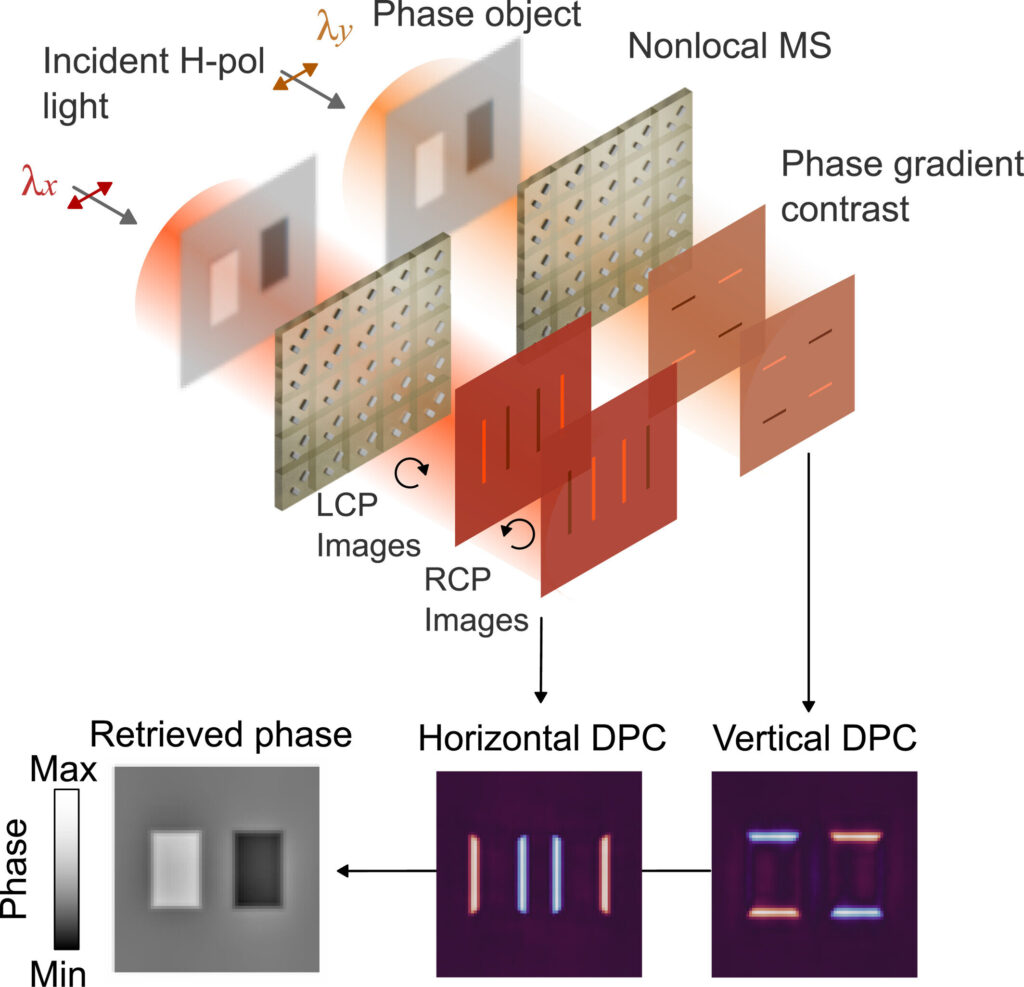
Full QPM requires measuring how the phase changes along perpendicular directions of an image. This means taking two separate images: one that shows how the phase changes from left to right and another showing its shift up and down across the image.
However, measuring the phase gradient across both directions poses time and convenience costs.
Between capturing both images, the metasurface would need to be rotated or a new surface inserted, requiring precise alignment and/or repeating a lengthy characterization process.
The researchers realized they could avoid this if they could incorporate the capability to make these two measurements into a single surface.
They realized that by engineering the nanoscopic structures of the metasurface to respond to only a very narrow band of light, they could construct two sets of structures on the surface, each sensitive to a different wavelength of light. This meant they could select how the metasurface functioned based on the wavelength of light used on their sample, removing the need to physically alter any setup to get both images.
This principle of multiplexing capabilities enabled the researchers to incorporate another technique, called differential phase contrast, which reduced noise in the final QPM images.
With multiple functions in a single chip, Wang and his fellow researchers had developed a method for achieving QPM in a highly compact form factor without physically moving components.
For Wang, this was a big step forward not only for the applications they hope for but also for metasurface research in general:
“These techniques have been used quite a lot, but not in the same way we have done it … Here, we are applying them to access different … functionalities, which hasn’t been done before.”
From microscopes to miniaturized sensors
With multiple functions in a single nanoscale surface, the potential for smaller devices and a streamlined imaging process could prove invaluable in diagnosing diseases such as sickle cell disease, various cancers and neurodegenerative diseases, which rely on observing cell shape and growth patterns.
When asked about the potential applications of this technology, Wang replied, “I think it could go into small devices you could take into point-of-care settings … Instead of having to lug around a giant microscope system, we can try to slot this into a smaller device, like a microscope on a smartphone …”
The more compact and time-efficient these imaging devices become, the easier it may be to diagnose diseases and collect data for biological, biotechnology and materials science research.
However, in the short term, there are still hurdles to overcome in the costs of supporting hardware and manufacturing, but for Wang, these new challenges are simply a matter of time and effort.
“Right now, we use an electron beam that scans the entire surface, whereas the commercial nanoimprint lithography takes one shot.”
When asked about the next step on the road to a true point-of-care system, Wang noted that “There is also a cost associated with generating the illumination … and it would still require a laser that can be tuned between two wavelengths very precisely.
“All the supporting components, right now, are implemented with traditional optics still.
“… Now I’m integrating our design into a commercial microscope … This one does not require a laser … It still needs an infrared LED, but this one is quite simple to insert into the system.”
When asked where this technology was ultimately headed, Wang said, “Miniaturized sensors are the main direction,” suggesting that as these sensors inevitably become smaller, they will see an expanding array of applications, from microscopic internal medicine to astronomy.
“People have been trying to put this on the head of an endoscope, which can be a bundle of fibers, and then you can move it inside the body. Then you can do imaging in vivo … that is really showing how something can be really miniaturized. The end of an endoscope is extremely small.
“… Another application is wavefront sensing. So, these are in a lot of telescopes to correct the effects of turbulence … Wavefront sensing is sort of similar to phase imaging.”
Wang was confident that this marked a new step for metasurface research in general, as their paper also concluded:
“To our knowledge, this is the first time such a multiplexing scheme has been demonstrated, and this work opens the path to exploring additional functionalities that can be multiplexed onto a single-layer metasurface.”
More information
Haiwei Wang et al, Wavelength and Polarization Multiplexed Nonlocal Metasurface for Quantitative Phase Microscopy, Nanophotonics (2026). DOI: 10.1002/nap2.70190
Clinical categories


Citation:
Two devices in one: Metasurface could bring medical imaging to smaller sensors (2026, August 15)
retrieved 15 August 2026
from https://medicalxpress.com/news/2026-08-devices-metasurface-medical-imaging-smaller.html
This document is subject to copyright. Apart from any fair dealing for the purpose of private study or research, no
part may be reproduced without the written permission. The content is provided for information purposes only.

Khamrah by Lattafa for Men - 3.4 oz EDP Spray
4% Off
Ghost Sweetheart Eau de Toilette | Pineapple, Jasmine and Sandalwood | Perfume for Women 50 ml
50% Off
Marc Jacobs Dot Eau De Parfum for Women, 100 ml
42% Off
Ted Baker W Eau de Toilette for Her, Fig Leaf, White Peony and Violet Top Notes, Pink Orchid and Raspberry Middle Notes, 75ml
£11.77 (£15.69 / 100 ml) (as of 10/09/2026 05:01 GMT +01:00 - More infoProduct prices and availability are accurate as of the date/time indicated and are subject to change. Any price and availability information displayed on [relevant Amazon Site(s), as applicable] at the time of purchase will apply to the purchase of this product.)
Ted Baker Woman Pink Eau de Toilette Spray Floral Green Feminine Fragrance, Opening Notes are Fresh Peach, Bergamot and Tangerine with Warm Musk, Vanilla and Vetiver Base, 100ml
11% Off
Vera Wang Princess Eau de Toilette - 30 ml

Choco Musk 50ml Eau De Parfum for men and women | Chocolate Musk by Jannat Aromas
17% Off
Christina Aguilera Signature Eau de Parfum (50ml) Floral, Fruity & Exotic Scent, Luxury Fragrance for Women
9% Off
Calvin Klein - Eau De Toilette CKIN2U - Calvin Klein Women, Ladies Perfume, Women's Perfume, Calvin Klein Perfume, Calvin Klein One - 150 ml
5% Off
Jimmy Choo Flash Eau de Parfum, 60 ml (Pack of 1)
3% Off
Fruit of the Loom Men's Heavy T Shirt, White, XL UK
28% Off
ATNKE LED Lighted Beanie Cap,USB Rechargeable Running Hat Ultra Bright 4 LED Waterproof Light Winter Warm Gifts for Men and Women/Pink
17% Off
Men's 1/4 Zip Pullover UK Sale Clearance, Fleece Sweatshirt Casual Jumper Long Sleeve T-shirt Top Stand Collar Sweater Plain Pullover Sports Leisure Workwear Quarter Zip Sweater Lightweight Jumpers
£5.88 (as of 12/11/2025 00:52 GMT +01:00 - More infoProduct prices and availability are accurate as of the date/time indicated and are subject to change. Any price and availability information displayed on [relevant Amazon Site(s), as applicable] at the time of purchase will apply to the purchase of this product.)
Crevice Cleaning Brush, Bathroom Tile Groove Gap Cleaning Brush,Premium Crevice Cleaning Tool Aluminum Support with 15° Angle Magic Brush, Thin Brush for Home Kitchen
19% Off
Wireless Earbuds, Bluetooth 5.3 Headphones in Ear with HiFi Stereo Deep Bass, 4 ENC Noise Cancelling Mic Wireless Earphones 40H Playtime, Bluetooth Earbuds Dual LED Display, IP7 Waterproof, USB-C
42% Off