 [
[


Major depressive disorder (MDD) is a common mental health condition primarily characterized by persistent low mood or loss of interest in daily activities. While several medications are available to treat depression, finding one that works for each patient can take time.
Understanding what happens in the brain shortly after treatment begins could help researchers identify how antidepressants work and, eventually, improve the treatment selection process.
Researchers at Stanford University School of Medicine, the University of Texas at Austin and other institutions recently analyzed brain scans from 386 people with MDD. They examined how connections between brain regions changed during the first one or two weeks after participants began receiving a placebo or one of two antidepressants. Their paper, published in Nature Mental Health, describes changes shared across treatment groups, as well as patterns more specific to the antidepressant medications examined.
“Although pretreatment brain signatures have been associated with MDD diagnosis and antidepressant response, the precise neural circuits underlying these associations remain elusive,” wrote Xiaoyu Tong, Gregory A. Fonzo and their colleagues in their paper. “To bridge this gap, we systematically characterize early treatment-induced changes in functional connectivity (FC) following 1 or 2 weeks of antidepressant or placebo administration using two independent cohorts of patients with MDD who are receiving medication.”
Tracking brain connections during early depression treatment
Tong, Fonzo and their colleagues analyzed resting-state functional magnetic resonance imaging (fMRI) scans collected from 386 adults with MDD ages 18 to 65. This type of scan measures changes in blood flow associated with brain activity while a person is resting. The researchers used the scans to study functional connectivity, or how closely activity in different brain areas rises and falls together.
Of the participants, 125 received a placebo (an inactive treatment), 123 received sertraline and 138 received escitalopram. Sertraline and escitalopram are widely prescribed selective serotonin reuptake inhibitors (SSRIs), antidepressants that increase the availability of the chemical messenger serotonin in the brain.
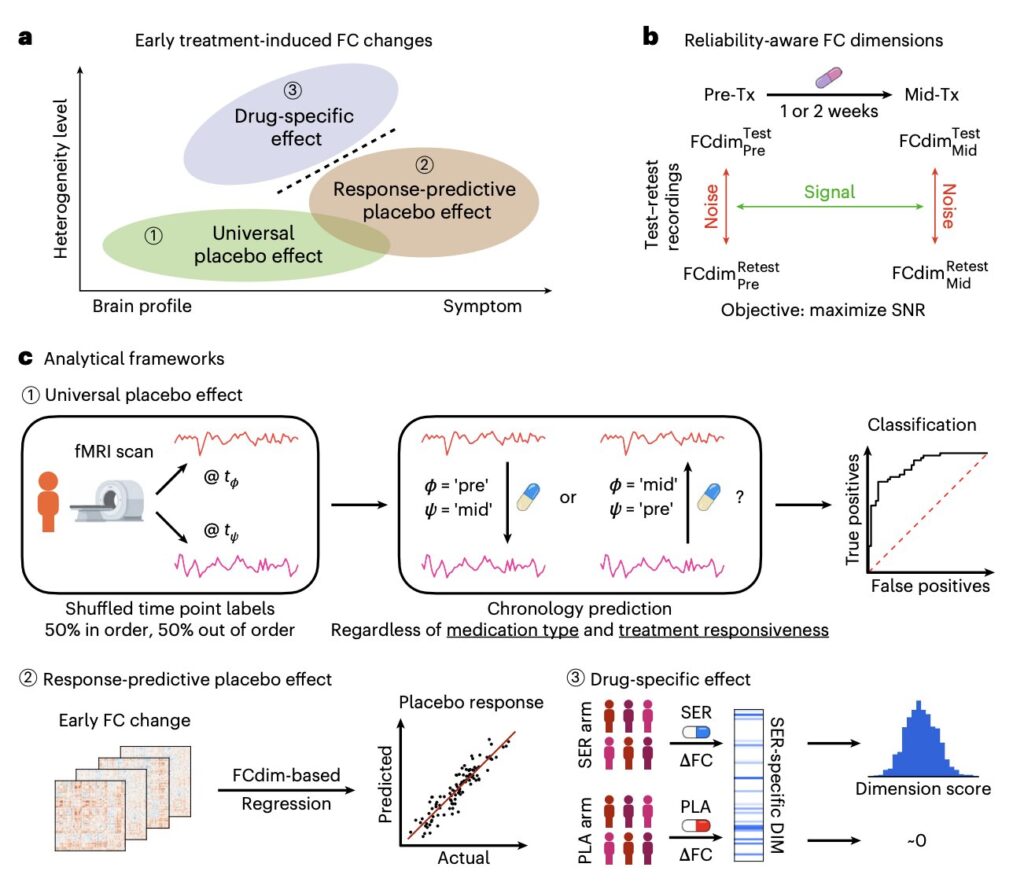
The team compared scans taken before treatment with scans taken one or two weeks after treatment began. Using computer models, they looked for changes in brain connections shared across participants, changes associated with symptom improvement in the placebo group and patterns more specific to treatment with the two antidepressants.
“Leveraging innovative predictive and contrastive machine learning frameworks, we identify a visual–precuneus–thalamus system exhibiting increased FC across patients, regardless of treatment or clinical outcome, and implicate striatal and attention networks in mediating placebo-related symptom improvement,” wrote the authors.
“Drug-specific effects center on the amygdala, midcingulate, orbitofrontal cortex and cerebellum but are present only in a subset of patients treated with antidepressants; notably, the responses of those without such changes can be predicted with a placebo response prediction model.”
Shared changes and drug-specific effects
The researchers identified an increase in functional connectivity within a system involving areas that process visual information, the precuneus (an area toward the back of the brain) and the thalamus (a relay center deep in the brain). These changes appeared across treatment groups, regardless of whether the participants’ symptoms had improved.
In the placebo group, symptom improvement was associated with changes involving the striatum, a brain region involved in motivation and reward, and networks involved in attention.
The team also identified functional connectivity patterns more specific to antidepressant treatment, which were observed in only a subset of participants taking the antidepressants. The changes involved the amygdala (a region that helps process emotionally significant information), the midcingulate and orbitofrontal cortices (areas of the brain’s outer layer), and the cerebellum (a region involved in movement and other functions).
“These findings reveal generalizable antidepressant-induced early FC changes, parse these changes into constituent placebo and drug-specific effects, and offer mechanistic insights into antidepressant action, supporting the development of interactive treatment optimization for MDD,” wrote the authors.
The results could inform further research into why patients respond differently to available depression treatments. Eventually, they could help clinicians plan treatment using brain scans collected shortly after a patient starts a new medication. However, more research is needed before the patterns identified in this study can guide clinical practice.
Written for you by our author Ingrid Fadelli, edited by Lisa Lock, and fact-checked and reviewed by Robert Egan—this article is the result of careful human work. We rely on readers like you to keep independent science journalism alive.
If this reporting matters to you, please consider a donation (especially monthly). You’ll get an ad-free account as a thank-you.
Publication details
Xiaoyu Tong et al, Early brain functional connectivity changes induced by antidepressants and placebo, Nature Mental Health (2026). DOI: 10.1038/s44220-026-00729-y
Journal information:
Nature Mental Health


© 2026 Science X Network
Citation:
Brain scans show shared changes during depression treatment and distinct antidepressant effects in some patients (2026, October 3)
retrieved 3 October 2026
from https://medicalxpress.com/news/2026-09-brain-scans-depression-treatment-distinct.html
This document is subject to copyright. Apart from any fair dealing for the purpose of private study or research, no
part may be reproduced without the written permission. The content is provided for information purposes only.

Khamrah by Lattafa for Men - 3.4 oz EDP Spray
4% Off
Ghost Sweetheart Eau de Toilette | Pineapple, Jasmine and Sandalwood | Perfume for Women 50 ml
50% Off
Marc Jacobs Dot Eau De Parfum for Women, 100 ml
42% Off
Ted Baker W Eau de Toilette for Her, Fig Leaf, White Peony and Violet Top Notes, Pink Orchid and Raspberry Middle Notes, 75ml
£11.77 (£15.69 / 100 ml) (as of 04/10/2026 05:09 GMT +01:00 - More infoProduct prices and availability are accurate as of the date/time indicated and are subject to change. Any price and availability information displayed on [relevant Amazon Site(s), as applicable] at the time of purchase will apply to the purchase of this product.)
Ted Baker Woman Pink Eau de Toilette Spray Floral Green Feminine Fragrance, Opening Notes are Fresh Peach, Bergamot and Tangerine with Warm Musk, Vanilla and Vetiver Base, 100ml
11% Off
Vera Wang Princess Eau de Toilette - 30 ml

Choco Musk 50ml Eau De Parfum for men and women | Chocolate Musk by Jannat Aromas
17% Off
Christina Aguilera Signature Eau de Parfum (50ml) Floral, Fruity & Exotic Scent, Luxury Fragrance for Women
9% Off
Calvin Klein - Eau De Toilette CKIN2U - Calvin Klein Women, Ladies Perfume, Women's Perfume, Calvin Klein Perfume, Calvin Klein One - 150 ml
5% Off
Jimmy Choo Flash Eau de Parfum, 60 ml (Pack of 1)
3% Off
Fruit of the Loom Men's Heavy T Shirt, White, XL UK
28% Off
ATNKE LED Lighted Beanie Cap,USB Rechargeable Running Hat Ultra Bright 4 LED Waterproof Light Winter Warm Gifts for Men and Women/Pink
17% Off
Men's 1/4 Zip Pullover UK Sale Clearance, Fleece Sweatshirt Casual Jumper Long Sleeve T-shirt Top Stand Collar Sweater Plain Pullover Sports Leisure Workwear Quarter Zip Sweater Lightweight Jumpers
£5.88 (as of 12/11/2025 00:52 GMT +01:00 - More infoProduct prices and availability are accurate as of the date/time indicated and are subject to change. Any price and availability information displayed on [relevant Amazon Site(s), as applicable] at the time of purchase will apply to the purchase of this product.)
Crevice Cleaning Brush, Bathroom Tile Groove Gap Cleaning Brush,Premium Crevice Cleaning Tool Aluminum Support with 15° Angle Magic Brush, Thin Brush for Home Kitchen
19% Off
Wireless Earbuds, Bluetooth 5.3 Headphones in Ear with HiFi Stereo Deep Bass, 4 ENC Noise Cancelling Mic Wireless Earphones 40H Playtime, Bluetooth Earbuds Dual LED Display, IP7 Waterproof, USB-C
42% Off